Download
1 / 31
310 likes | 438 Views
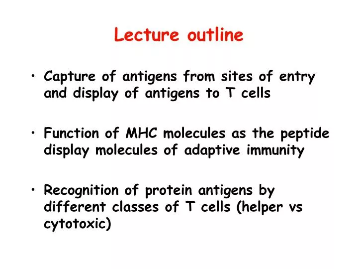
Lecture outline . Capture of antigens from sites of entry and display of antigens to T cells Function of MHC molecules as the peptide display molecules of adaptive immunity Recognition of protein antigens by different classes of T cells (helper vs cytotoxic) . Functions of T lymphocytes.

E N D
Lecture outline • Capture of antigens from sites of entry and display of antigens to T cells • Function of MHC molecules as the peptide display molecules of adaptive immunity • Recognition of protein antigens by different classes of T cells (helper vs cytotoxic)
Functions of T lymphocytes • Defense against cell-associated microbes • Helper T cells help phagocytes to kill ingested microbes and help B cells to make potent antibodies • Cytotoxic T lymphocytes (CTLs) kill infected cells and eliminate reservoirs of infection • Inhibition of immune responses • Regulatory T cells suppress APCs or other lymphocytes • T cell functions require cell-cell interactions or cytokines that act at short range
The challenge for T lymphocytes • Very few lymphocytes in the body are specific for any one microbe (or antigen) • Specificity and diversity of antigen receptors: the immune system recognizes and distinguishes between 106 - 109 antigens; the body contains ~ 1012 lymphocytes; therefore, few lymphocytes (~1,000) can recognize any one antigen and need to find that antigen
The challenge for T lymphocytes • Very few lymphocytes in the body are specific for any one microbe (or antigen) • Specificity and diversity of antigen receptors: the immune system recognizes and distinguishes between 106 - 109 antigens; therefore, few lymphocytes can recognize any one antigen and need to find that antigen • Lymphocytes must be able to locate and respond to microbes that enter and reside anywhere in the body • Usual routes of entry are through epithelia, but infections may take hold anywhere • It is not possible for lymphocytes of all specificities to patrol all the tissues where antigens may be located
The challenge for T lymphocytes • Very few lymphocytes in the body are specific for any one microbe (or antigen) • Specificity and diversity of antigen receptors • Lymphocytes must be able to locate microbes that enter anywhere • Usual routes of entry are through epithelia • Lymphocytes must respond to each microbe in ways that are best able to eradicate that microbe • Response to extracellular microbes: antibodies that promote phagocytosis; destruction in macrophages (need helper T cells) • Response to intracellular microbes: killing of infected cells (need CTLs) • Cannot be based on different specificities of helper cells and CTLs
Antigen capture Sites of antigen entry Sites of initial antigen capture Sites of antigen collection and capture
Capture and presentation of antigens by dendritic cells Sites of microbe entry: skin, GI tract, airways (organs with continuous epithelia, populated with dendritic cells). Less often -- colonized tissues, blood Sites of lymphocyte activation: peripheral lymphoid organs (lymph nodes, spleen), mucosal and cutaneous lymphoid tissues) Antigens and naïve T cells come together in lymphoid organs
Why are dendritic cells the most efficient APCs for initiating immune responses? • Location:at sites of microbe entry (epithelia), tissues • Receptors for capturing and reacting to microbes:Toll-like receptors, mannose receptors, others • Migration to T cell zones of lymphoid organs • Role of CCR7 • Co-localize with naïve T cells • Maturation during migration:Conversion from cells for antigen capture into cells for antigen presentation and T cell activation • Practical application:dendritic cell-based vaccines for cancer immunotherapy
What do T cells see? • All functions of T cells are mediated by interactions with other cells • Helper T cells “help” B cells to make antibodies and “help” macrophages to destroy what they have eaten • Cytotoxic (killer) T lymphocytes kill infected cells • How does the immune system ensure that T cells see only antigens on other cells?
What do T cells see? • All functions of T cells are mediated by interactions with other cells • Helper T cells “help” B cells to make antibodies and “help” macrophages to destroy what they have eaten • Cytotoxic (killer) T lymphocytes kill infected cells • To ensure cellular communications, T cells see antigens NOT in the circulation but only when displayed by molecules on the surface of other cells • These molecules are the MHC (HLA) molecules, and the cells displaying the antigen are APCs
Schematic model of T cell recognition of antigen During their maturation in the thymus, T cells whose TCRs see MHC molecules are selected to mature, i.e. mature T cells are MHC-restricted
What is the MHC? • A genetic locus discovered on the basis of transplantation (major histocompatibility complex) • Different individuals express products of different MHC alleles and reject grafts from one another • Human MHC: HLA (human leukocyte antigens) • MHC molecules are the peptide display molecules of the immune system
What is the MHC? • A genetic locus discovered on the basis of transplantation (major histocompatibility complex) • Different individuals express products of different MHC alleles and reject grafts from one another • Human MHC: HLA (human leukocyte antigens) • MHC molecules are the peptide display molecules of the immune system • Different alleles of MHC molecules bind and display distinct but overlapping sets of peptides • Determines which protein antigens are recognized in different individuals • MHC genes are highly polymorphic (>7500 alleles [variants] in the population); the MHC molecules in the population can display many different peptides
What is the MHC? • A genetic locus discovered on the basis of transplantation (major histocompatibility complex) • Different individuals express products of different MHC alleles and reject grafts from one another • Human MHC: HLA (human leukocyte antigens) • MHC molecules are the peptide display molecules of the immune system • Different alleles of MHC molecules bind and display distinct but overlapping sets of peptides • Determines which protein antigens are recognized in different individuals • MHC genes are highly polymorphic; the MHC molecules in the population can display many different peptides • MHC molecules determine how antigens in different cellular compartments are recognized by different classes of T cells (CD4+ and CD8+)
MHC-restricted antigen recognition by T cells • Any T cell can recognize an antigen on an APC only if that antigen is displayed by MHC molecules • Antigen receptors of T cells have dual specificities: 1. for peptide antigen (responsible for specificity of immune response) and 2. for MHC molecules (responsible for MHC restriction) • During maturation in the thymus, T cells whose antigen receptors see MHC are selected to survive and mature; therefore, mature T cells are “MHC-restricted”
The genes of the MHC locus Genes in the MHC locus encode most of the proteins that form the machinery of antigen processing and presentation
peptide binds CD4 binds CD8 All MHC molecules have a similar basic structure: the tops bind peptide antigens and are recognized by T cell receptors and the “tails” bind CD4 or CD8.
Antigen processing • Conversion of native antigen (large globular protein) into peptides capable of binding to MHC molecules • Occurs in cellular compartments where MHC molecules are synthesized and assembled • Determines how antigen in different cellular compartments generates peptides that are displayed by class I or class II MHC molecules
Pathways of antigen processing Protein antigen in vesicles --> class II MHC pathway Protein antigen in cytosol (cytoplasm) --> class I MHC pathway
The class II MHC pathway of processing of internalized vesicular protein antigens Endocytosed proteins are cleaved into peptides in vesicles; class II MHC molecules are available to bind the peptides in the same vesicles
Class II MHC pathway of presentation of vesicular peptide antigens • Helper T cells need to help macrophages and B cells that have encountered (and ingested) microbes • Proteins ingested into endosomes/lysosomes (vesicles) are processed and their peptides are presented in association with class II MHC molecules • Most vesicular peptides are derived from extracellular proteins that are ingested into vesicles • Class II MHC is expressed only on specialized cells (e.g. B cells, macrophages) that are capable of ingesting microbes and antigens into vesicles
Class II MHC pathway of presentation of vesicular peptide antigens • Proteins ingested into endosomes/lysosomes are processed and their peptides arepresented in association with class II molecules • Most vesicular peptides are derived from extracellular proteins that are ingested • Class II MHC is expressed only on specialized cells (e.g. B cells, macrophages) that are capable of ingesting microbes and antigens into vesicles • CD4 binds to class II MHC; therefore, CD4+ T cells recognize class II-displayed peptides • CD4+ T cells are helper cells that activate B lymphocytes and macrophages • Antibodies (products of activated B cells) and activated macrophages combat extracellular microbes
The class I MHC pathway of processing of endogenous cytosolic protein antigens Cytoplasmic peptides are actively transported into the ER; class I MHC molecules are available to bind peptides in the ER
Class I MHC pathway of presentation of cytosolic peptide antigens • Cytotoxic T lymphocytes need to kill cells containing cytoplasmic microbes, and tumor cells (which contain tumor antigens in the cytoplasm) • Cytosolic proteins are processed into peptides that are presented in association with class I molecules • Most cytosolic peptides are derived from endogenously synthesized (e.g. viral, tumor) proteins • All nucleated cells (which are capable of being infected by viruses or transformed) express class I
Class I MHC pathway of presentation of cytosolic peptide antigens • Cytosolic proteins are processed into peptides that are presented in association with class I molecules • Most cytosolic peptides are derived from endogenously synthesized (e.g. viral, tumor) proteins; all nucleated cells (which are capable of being infected by viruses or transformed) express class I • CD8 binds to class I MHC; therefore, CD8+ T cells recognize class I-displayed peptides • CD8+ T cells are cytotoxic cells that kill any nucleated cells that harbor infections (thus eliminating reservoirs of infection) or are transformed
How class I- and class II-associated antigen presentation influences the nature of the T cell response
Cross presentation of microbial antigens from infected cells to CD8+ T cells Antigens of viruses or tumors that are produced in cells other than APCs have to elicit CTL responses, which usually requires DCs; antigen-producing cell is phagocytosed by DCs, and phagocytosed antigen enters the class I pathway (exception to the rule!)
Functions of APCs • Capture antigens and take them to the “correct” anatomic site • Antigens are concentrated in peripheral lymphoid organs, through which naïve lymphocytes circulate • Display antigens in a form that can be recognized by specific lymphocytes • For T cells: MHC-associated peptides (cytosolic peptides to class I, vesicular peptides to class II) • For B cells: native antigens • Provide “second signals” for T cell activation
APCs and self antigens • Normally, APCs are constantly presenting self antigens • MHC molecules do not distinguish self from foreign • If MHC molecules are bathed in self peptides, how can they ever be free to present microbial peptides? • Very few peptides (complexed with MHC) are enough to activate specific T cells • Microbes induce “second signals” on APCs • If self peptides are always being displayed, why do we not react against our own antigens?
How do T lymphocytes meet their challenges? • Very few lymphocytes in the body are specific for any one microbe (or antigen) • Lymphocytes must be able to locate and respond to microbes that enter anywhere in the body • Antigens are transported to and concentrated in the lymphoid organs through which naïve T cells constantly circulate, increasing likelihood of encounter • Lymphoid organs, which are specialized to initiate immune responses, drain all tissue sites
How do T lymphocytes meet their challenges? • Very few lymphocytes in the body are specific for any one microbe (or antigen) • Lymphocytes must be able to locate and respond to microbes that enter anywhere in the body • Lymphocytes must respond to each microbe in ways that are best able to eradicate that microbe • Antigens of endogenous and extracellular microbes are displayed to different subsets of T cells by class I and class II MHC molecules • Even the same microbe may be recognized by CD4+ or CD8+ T cells depending on its cellular location