Download
1 / 57
600 likes | 983 Views
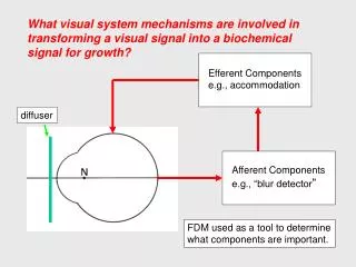
What visual system mechanisms are involved in transforming a visual signal into a biochemical signal for growth?. Efferent Components e.g., accommodation. diffuser. Afferent Components e.g., “blur detector ”. FDM used as a tool to determine what components are important. FDM in primates.

E N D
What visual system mechanisms are involved in transforming a visual signal into a biochemical signal for growth? Efferent Components e.g., accommodation diffuser Afferent Components e.g., “blur detector” FDM used as a tool to determine what components are important.
FDM inprimates FDM does NOT require: - the visual signal to leave the eye - sympathetic or parasympathetic inputs to the eye.
Restricted Form Deprivation Selectively depriving a portion of the eye restricts the axial elongation and myopia to the deprived areas. Wallman et al. 1978
Local Retinal Mechanisms Efferent Afferent The mechanisms that mediate the effects of visual experience on eye growth are located largely within the eye. Activity at a given retinal location controls the growth of the adjacent sclera.
Emmetropization Model (in mammals) Norton, 1999 Key points: 1. Ocular growth regulated by retinal responses to optical image. 2. Accommodation, by its influence on retinal image quality, plays an indirect role in emmetropization.
Acetylcholine (M1 or M4 receptors) • Dopamine (Acs) • Gulcagon (Acs) • Vasoactive Intestinal Peptide (Acs) • Nicotine (Antagonist effects) Retinal Components Norton, 1999
Effects of Chronic Atropine Chronic atropinization produces hyperopia in young monkeys and has been reported to slow the progression of myopia in humans.
Atropine and FDM Chronic atropinization prevents FDM in some species of monkeys.
Neurochemical Transmission in the Parasympathetic System Atropine produces cycloplegia by blocking the action of acetylcholine on muscarinic receptors in ciliary muscle.
Cholinergic Receptor Subtypes M1 CNS, nerves M2 Heart, smooth muscle, ciliary muscle M3 Smooth muscle, exocrine glands, ciliary muscle M4 CNS, nerves M5 CNS, ciliary muscle Atropine blocks all muscarinic receptor subtypes.
Blocking actions: atropine - all muscarinic sites 4-DAMP - smooth muscle pirenzepine - neural ganglia
Tree Shrew: Pirenzepine & FDM Atropine and pirenzepine are effective in preventing FDM in tree shrews. Other selective muscarinic antagonists (M2, gallamine; M3, P-f-HHSid) were not effective in blocking FDM. Hence, the M1 receptor appears to have potential therapeutic value. M1 blockers do not eliminate accommodation. McBrien et al., 2000
Activity Markers in Amacrine Cells Glucagon amacrine cells are more abundant than dopaminergic Acs. Tested for visual regulation of several transcription factors. Conditions that stimulate axial elongation decrease ZENK synthesis (basically glucagon activity) whereas conditions that reduce axial growth up-regulate ZENK. Glucagon AC exhibit sign of defocus information. Seltner & Stell, 1995
Choroidal Components • Choroidal Retinoic Acid • Choroidal Thickness Norton, 1999
Choroidal Retinoic Acid Synthesis: Mediator of Eye Growth? Evidence in chicks: 1) the choroid can convert retinol to all-trans retinoic acid at a rapid rate. 2) Visual conditions that increase ocular growth produce a sharp decrease in retinoic acid synthesis. 3) Visual conditions that slow ocular growth produce an increase in RA synthesis. 4) application of RA to cultured sclera inhibits proteoglycan production at physiological concentrations. Mertz et al., 2000a
Choroidal Mechanisms Changes in choroid thickness move the retina toward the appropriate focal point. chick recovering from induced myopia normal chick from Wallman et al., 1995
Scleral Components • bFGF & TGF beta (growth factors) • For a myopic stimulus: • Decrease proteoglycan synthesis • Decrease sulfated GAGs • Increase gelatinolytic enzymes Norton, 1999
Possible growth factors involved in FDM bFGF = basic fibroblast growth factor. TGF-beta = transforming growth factor beta. The broad dose response curve suggests that more than one type of FGF receptor is involved.
Scleral Changes with FDM Matrix metalloproteinase (MMP-2) appears to be the major gelatinolytic enzyme in the tree shrew sclera. Form deprivation increases catabolism in the sclera. Hyperopic defocus reduces the degree of scleral catabolism. Guggenheim & McBrien, 1996
Scleral Changes with FDM Rada et al., 2000 Decorin is the major proteoglycan in the marmoset sclera. The rate of proteoglycan synthesis is reduced in the posterior pole of FDM.
Physical Changes Norton, 1999 • Increase / decrease in scleral creep rate • Axial vitreous chamber depth
Scleral Changes with FDM The scleras from eyes that are undergoing myopic axial elongation exhibit higher than normal creep rates. During recovery from FDM the scleral creep rates fell below normal values. During both emmetropization and the development of refractive errors, vision-dependent alterations in the extracellular matrix may alter the mechanical properties of the fibrous sclera making it more distensible. Siegwart & Norton, 1995
Why Worry About Myopia? • Myopia is common. • 36% of all prescriptions in USA. • Myopia is expensive. • Total direct costs ($ billions) – estimated for 2000 in USA • $5 to $6 Spectacles & contact lenses • $1.6 to $1.9 Professional Services • $2.2 Refractive Surgery • Inconvenience and complications of correcting strategies.
Ocular Sequelae of Myopia Posterior Subcapsular Cataract 2 to 5 X Open-Angle Glaucoma 2.2 X Idiopathic Retinal Detachment 4 to 10 X (Curtin, 1985) Chorioretinal Degeneration
The idea that something about near work causes myopia has dominated thinking for centuries. Duke-Elder, 1970 • Theoretical basis for traditional therapy • - Increased IOP • - Excessive convergence &/or accommodation • Gravity & posture Levinson, 1919
Lag of Accommodation Myopic children accommodate significantly less than emmetropic children for real targets at near distances. Gwiazda et al, 1993
Do bifocals reduce the rate of myopic progression? Investigative Ophthalmology & Vision Research, September 2002 Randomized, double-masked clinical trial to determine whether progressive addition lenses (SOLA MC lenses with a near addition of +1.50 D) reduce the progression of myopia in children over a 2 year period.
Longitudinal Changes in Refractive Error and Axial Length Mean ± SEM Edwards et al., 2002 At the end of the treatment period, the PAL group was on average 0.25 D less myopic.
The Comet Study Investigative Ophthalmology & Vision Science 44:1492, 2003 Randomized, double-masked clinical trial to determine whether progressive addition lenses (Varilux Comfort Lenses with a near addition of +2.00D) reduce the progression of myopia in children over a 3 year period.
The Comet Study Myopic Progression PALs SV Gwiazda et al., 2003
The Comet Study PALs reduce progression rate by about 50% (about 0.75 D in 3 years) in esophores with large lags of accommodation. Gwiazda et al., 2004
Do Near Adds Eliminate Accommodative Errors? Optimal Add? Subjects typically fail to relax accommodation by an amount equal to the add. Near adds may actually increase the degree of retinal defocus. Rosenfield & Carrel, 2001
Does undercorrection slow myopic progression? Randomized, controlled clinical trial to determine the effects of undercorrection on the rate of progression of myopia.
MethodsSubject Selection Criteria • Age: 9-14 years. • At least –0.5 D of myopia (sph equiv) in both eyes & myopic in all meridians. • < 2.0 D of astigmatism in each eye. • Corrected VA = 20/20 or better in each eye. • No significant binocular vision problems. • Normal ocular health. • No previous contact lens wear.
Methods Chung, Mohidin and O’Leary • Spectacle Corrections: • Full Correction: Maximum plus to obtain best VA in each eye. Full compliance 41 of 46. • Undercorrection: Monocular VA maintained at 20/40 by undercorrecting by about +0.75 D. Full compliance 40 of 47. • Patients instructed to wear spectacles at all times. Full Compliance > 8 hours/day.
Mean Changes in Refractive Error The undercorrected group showed a greater rate of myopic progression. Fully Corrected Start of Trial Undercorrected Average sph equivalent (± SEM) for both eyes. From Chung et al., 2002
Mean Changes in Axial Length The undercorrected group showed greater axial elongation. Undercorrected Fully Corrected No between group differences in corneal curvature, anterior chamber depth or lens thickness. Start of Trial From Chung et al., 2002
The “CLAMP” Study Contact Lens and Myopia Progression RGPS vs Soft CLs Walline et al., 2004
The “CLAMP” Study Walline et al., 2004
The “CLAMP” Study Walline et al., 2004
New Hopes for Optical Interventions Emmetropization: Basic Operating Properties
Visual Signals for Axial Growth Ferree & Rand, 1933 Central vs. 30 deg Nasal Refractive error varies with eccentricity. Myopes typically exhibit relative hyperopia in the periphery, whereas hyperopes show relative myopia in the periphery. Mutti et al., 2000
Should we correct peripheral refractive errors? As a consequence of eye shape and/or aspheric optical surfaces, myopic eyes may experience significant defocus across the visual field, regardless of the refractive state at the fovea. Uncorrected Myope Image Shell “Corrected” Myope Optimal Correction?
Timolol Treatment for Myopia Timolol was effective in lowering IOP. However there was not a significant effect on the rate of myopic progression. Jensen, 1991
Schmidt & Wildsoet, 2000 Timolol and Form-Deprivation Myopia Timolol was effective in lowering IOP in young chicks (between 18 & 27%). However there was not a significant effect on the rate of myopic progression for either form deprivation or negative lenses