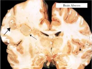
Brain Abscess
Brain Abscess. Microorgansims reach the brain by i. Direct extension ii. Hematogenous spread Iii. Direct inoculation from penetrating trauma or neurosurgical intervention. Brain Abscess. Younger patients affected (<40 years) Presence of predisposing condition in 80% of cases

Brain Abscess
E N D
Presentation Transcript
Brain Abscess • Microorgansims reach the brain by • i. Direct extension • ii. Hematogenous spread • Iii. Direct inoculation from penetrating trauma or neurosurgical intervention
Brain Abscess • Younger patients affected (<40 years) • Presence of predisposing condition in 80% of cases • Immunocompromised states from AIDS and immunosuppressive drugs in organ transplant recipents
Most Common Pathogens • Otitis media, mastoiditis Streptococci • Paranasal sinusitis Streptococci • Pulmonary infection Strep, Actionomyces • Dental Mixed, Bacteroides spp. • CHD Strep • Penetrating/Post-crani S. aureus • HIV Toxoplasma gondii • Transplant Aspergillus, Candida
Treatment • I.V. Antibiotics 6 weeks • Steroids • Surgical intervention: Stereotactic aspiration vs. craniotomy
PITUITARY APOPLEXY Clinical Scenario Jan M. Eckermann, MD Department of Neurosurgery
Objectives • Definition • Anatomy and Physiology • Pathophysiology • Signs and Symptoms • Differential diagnosis • Treatment • Prognosis and Outcomes
Definition • Clinical syndrome characterized by sudden headache, vomiting, visual impairment and meningismus caused by rapid enlargement of a pituitary adenoma usually due to hemorrhagic infarction of the tumor • Pituitary apoplexy is a clinical definition
Incidence • 0.6 – 9.1% apoplexy in pituitary adenomas treated surgically • 0.6 – 25.7% hemorrhage in pituitary adenomas treated surgically • Male: Female: 1.3:1 • Mean age: 46.7 years
Pathophysiology • Controversial • Rapid growth of tumor outstrips blood supply, producing ischemic necrosis hemorrhage • Direct invasion of vessel wall by tumor and consequent vessel rupture • Differences in vasculature of adenoma and normal adenohypophesis
Pathophysiology • Compromised blood flow caused by compression of pituitary stalk • High pressure system through inferior hypophyseal arteries causes hemorrhages in low-pressure adenohypophyseal sinusoids • Increased intrasellar pressure (fragile neovascularization)
Pathophysiology • Null-cell adenomas have highest incidence of apoplexy • Size, apparently, does not matter • Most cases show necrosis, hemorrhage, or both • Pituitary apoplexy as been described in association with a variety of conditions • Most common predisposing factor, however unproven, is arterial hypertension
Signs and Symptoms • Headache 100% (often retro-orbital) • Nausea 80% • Reduction in visual field 71% • Ocular paresis 69% • Third nerve palsy 67% • Reduction in visual acuity 66% • Vomiting 57% • Photophobia 49% • Decreased level of consciousness 11%
Investigations • Biochemical: • Gonadotropin deficiency 79% • Hypocortisolism 76% • Testosterone deficiency 73% • TSH deficiency 50% • Hyponatremia (<135) 44%
Investigations • Radiological: • CT scan revealed tumor in 93% and hemorrhage in 21% • MRI revealed tumor in 100% and hemorrhage in 88%
Differential Diagnosis • SAH from aneurysmal rupture • Spontaneous hemorrhage from hypertension, amyloid angiopathy • Migraine • Temporal arteritis • Meningitis • Diabetic oculomotor palsy • Optic neuritis • Cavernous sinus thrombosis
Treatment • Management focused on two aspects: i. Endocrinopathy ii. Acute neurologic deficits from tumor mass
Treatment • Medical stabilisation • High-dose steroids • Pituitary panel and electrolytes • Imaging • Emergent surgical decompression • Endocrinologic consultation
Outcome and Prognosis • Lethal outcome very infrequent • Emergent decompression may recover pituitary function • Visual outcome: early decompression (<8 days) improves visual acuity and visual fields. No influence on ocular paresis (86%, 76%, 91%)
Outcome and Prognosis • Endocrinologic outcome: long-term replacement therapy in 43-58%, transient diabetes insipidus in 16%
The Bottom Line • Rapid, thorough evaluation • Pituitary panel • High-dose steroids • MRI • Unless patient presents with rapidly progressive visual or neurologic deficit, urgent but not emergent intervention is recommended.
References • Andrews Brian T. Intensive Care in Neurosurgery. Thieme: New York 2003 • Krisht AF and Tindall GT. Pituitary Disorders Comprehensive Management. Lippincott Williams and Wilkins: Baltimore 1999 • Randeva HS, Schoebel J, Byrne J, et al. Classical Pituitary Apoplexy: Clinical Features, Management and Outcome. Clinical Endocrinology (1999) 51, 181-188 • Rengachary SS and Ellenbogen RG. Principles of Neurosurgery 2nd Edition. Elsevier Mosby: Edinburgh 2005 • Stein JH. Internal Medicine Fifth Edition. Mosby: St. Louis 1998