Download
1 / 117
1.18k likes | 1.36k Views
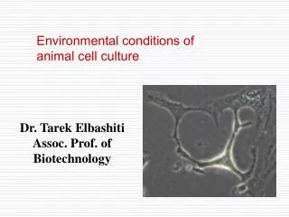
Biology of cultured cells. Dr. Tarek Elbashiti Assoc. Prof. of Biotechnology. In vitro cell often does not express the correct in vivo phenotype because the cell’s microenvironment has changed.

E N D
Biology of cultured cells Dr. Tarek Elbashiti Assoc. Prof. of Biotechnology
In vitro cell often does not express the correct in vivo phenotype because the cell’s microenvironment has changed. Cell–cell and cell–matrix interactions are reduced because the cells lack the heterogeneity and three-dimensional architecture found in vivo, and many hormonal and nutritional stimuli are absent.
The influence of the environment on the culture is expressed via five routes: (1) the nature of the substrate on or in which the cells grow—solid, as on plastic or other rigid matrix, semisolid, as in a gel such as collagen or agar, or liquid, as in a suspension culture (2) the degree of contact with other cells (3) the physicochemical and physiological constitution of the medium (4) the constitution of the gas phase (5) the incubation temperature.
The providing of the appropriate environment, including substrate adhesion, nutrient and hormone or growth factor concentration, and cell interaction, is fundamental to the expression of specialized functions
Cell adhesion Most cells from solid tissues grow as adherent monolayers, and, unless they have transformed and, Become anchorage independent after tissue disaggregation or subculture they will need to attach and spread out on the substrate before they will start to proliferate.
Cells attach to and spread on glass that had a slight net negative charge. • Attach to some plastics, such as polystyrene, if treated with an electric ion discharge or high-energy ionizing radiation. • Cell adhesion is mediated by specific cell surface receptors for molecules in the extracellular matrix so it seems likely that spreading may be preceded by the secretion of extracellular matrix proteins and proteoglycans by the cells.
Cell Adhesion Molecules • Three major classes of transmembrane proteins have shown to be involved in cell-cell and cell-substrate adhesion: • Cell-cell adhesion molecules, CAMs (Ca2+ independent), and cadherins (Ca2+ dependent) for interactions between homologous cells. • Cell-substrate interactions by integrins, receptors for matrix molecules such as fibronectin, laminin, and collagen. • Transmembrane proteoglycans, also interacting with matrix such as other proteoglycans or collagen
CELL JUNCTIONS Adherens junctions Gap junctions Tight junctions Desmosomes/Hemidesmosomes Focal adhesions
Intercellular Junctions • The role of the junctions varies between mechanical, such as the desmosomes and adherens junctions, which hold epithelial cells together • Tight junctions which seal the space between cells, e.g. between secretory cells in ducts or between endothelial cells in a blood vessel, and • Gap junctions, which allow ions, nutrients, and small signaling molecules such as cyclic adenosine monophosphate (cAMP) to pass between cells in contact.
Desmosomes distributed throughout the area of plasma membranes in contact they are often associated with tight and adherens junctions. • Desmosomes are molecular complexes of cell adhesion proteins and linking proteins that attach the cell surface adhesion proteins to intracellular keratin cytoskeletal filaments.
As epithelial cells differentiate in confluent cultures they can form an increasing number of desmosomes and, if some morphological organization occurs, can form complete junctional complexes.
This is one reason why epithelial cells, if left at confluence for too long, can be difficult to disaggregate. • As many of the adhesion molecules within these junctions depend on Ca2+ ions, a chelating agent, such as EDTA, is often added to the trypsin during or before disaggregation.
Extracellular Matrix • Intercellular spaces in tissues are filled with extracellular matrix (ECM), the constitution of which is determined by the cell type, e.g., fibrocytes secrete type I collagen and fibronectin into the matrix, • Epithelial cells produce laminin. • Where adjacent cell types are different, e.g., at the boundary of the dermis (fibrocytes) and epidermis (epithelial keratinocytes), both cell types contribute to the composition of the ECM, often producing a basal lamina.
The matrix adheres to the charged substrate, and the cells then bind to the matrix via specific receptors. • Glass or plastic that has been conditioned by previous cell growth can often provide a better surface for attachment, • Substrates pretreated with matrix constituents, such as fibronectin or collagen, or derivatives, such as gelatin, help fastidious cells to attach and proliferate.
Mostly, cultured cell lines are allowed to generate their own ECM, • but primary culture and propagation of some specialized cells, and the induction of their differentiation, may require exogenous condition of ECM. • ECM is comprised variously of collagen, laminin, fibronectin, hyaluronan, proteoglycans, and bound growth factors or cytokines.
With fibroblast-like cells, the main requirement is for substrate attachment and spreading and the cells migrate individually at low densities. • Epithelial cells may also require cell–cell adhesion for optimum survival and growth and, as a result, they tend to grow in patches.
At least two components of interaction with the substrate may be recognized: (1) Adhesion, to allow the attachment and spreading that are necessary for cell proliferation. (2) Specific interactions, such as of the interaction of an epithelial cell with basement membrane(a dense layer of extracellular material) with other ECM constituents, or with adjacent tissue cells, and required for the expression of some specialized functions and others explored the growth of cells on other natural substrates related to basement membrane.
The use of ECM constituents can be highly beneficial in enhancing cell survival, proliferation, or differentiation • Fibronectin and laminin fragments are now available commercially
CELL-CELL ADHESION MOLECULES Cadherins Ig superfamily CAMs Selectins Integrins Connexins (Gap Junction molecules) Occludin and claudin proteins
CELL-CELL ADHESION MOLECULES Transmembrane proteins involved in cell–cell and cell–substrate adhesion 1- (a) Cell–cell adhesion molecules, CAMs (Ca2+independent), by means of cell adhesion molecules, CAMs, cells are capable of recognizing each other Plasma membrane receptors take care of cell-ECM interactions (b) cadherins (Ca2+ dependent) primarily involved in interactions between homologous cells
2. Cell–substrate interactions are mediated primarily by integrins, receptors for matrix molecules such as fibronectin, entactin, laminin, and collagen, which bind to them via a specific motif usually containing the arginine–glycine–aspartic acid (RGD) sequence • Each integrin comprises one α and one β subunit, the extracellular domains of which are highly polymorphic, thus generating considerable diversity among the integrins.
3. The transmembrane proteoglycans, also interacting with matrix constituents such as other proteoglycans or collagen, but not via the RGD motif. • Disaggregation of the tissue, or an attached monolayer culture, with protease digest some of the extracellular matrix and may even degrade some of the extracellular domains of transmembrane proteins, allowing cells to become dissociated from each other.
Epithelial cells are generally more resistant to disaggregation, as they tend to have tighter junctional complexes (desmosomes, adherens junctions, and tight junctions) holding them together, whereas mesenchymal cells, which are more dependent on matrix interactions for intercellular bonding, are more easily dissociated. • Endothelial cells (specialized type of epithelial cell which forms the inner layer of blood vessels) may also express tight junctions in culture, especially if left at confluence for prolonged periods on a preformed matrix, and can be difficult to dissociate.
C.T: Functions to bind and support other tissues • Made up of a thin population of cells scattered through an extracellular matrix • Matrix – nonliving, web of fibers embedded in a homogenous ground substance that may be liquid, solid, or jelly-like • Substances of the matrix are secreted by cells of connective tissues • In each case, cells must re-synthesize matrix proteins before they attach or must be provided with a matrix-coated substrate.
The binding is homophilic Adhesion may be homophilic (one cadherin binds to another in the extracellular space andconnects cells together at specialized junctions)or heterophilic(binding protein binds to another type of site on a cell).
Cadherins Cadherins: Large family of cell surface proteins that mediate homophilic Ca++ dependent cell-cell adhesion. The classical cadherins, e.g. E-,N-, P-, L-cadherins occur in the epithelial, neuronal placental and liver tissues respectively.
700-750 a.a glycoprotein Five fold external parts Four with Ca++ In Ca++ deficiency !! Ca2+ causes dimerization of Cadherins
Cell-cell Adhesion Regulation of cadherins
CADHERINS They have a critical role in embryo morphogenesis. In adult, cadherins are responsible for the tight cell-cell associations within tissues. They are closely associated with the cytoskeleton – (actin ), By 3 intracellular proteins (catenins) Associated with signaling between the cell surface and the nucleus If catenins absent cadherin don’t act.
SELECTINS Surface carbohydrate binding protein. Ex. Lectins in the presence of Ca ions bind to specific oligosaccharides on another cell All are structurally closely related having, at their N-terminal a carbohydrate recognition (lectin) domain and variable numbers of repeats related to complement regulatory proteins. Heterophilic cell adhesion bind protein to another type on a cell. This binding is weak untill bind tightly by their integrins.
SELECTINS • There are three types of selectins • E-selectin, found exclusively on endothelia • L-selectin, found on all circulating leukocytes except activated T-lymphocytes • P-selectin, found in secretory granules of platelets and endothelial cells.
SELECTINS Selectins are involved in extravasations Inflammatory signals activate endothelial cells making P-Selectin undergo exocytosis P-Selectin on the surface of endothelial cells binds a specific carbohydrate ligand on leukocytes The leukocytes attach to the endothelial wall and roll slowly on it Platelet-activating factor (PAF) and integrins are then activated and the leukocytes start to passthrough the walls of a vessel into the surrounding tissues.
Integrins • Transmembrane binding glycoproteins that usually bind cells to matrix • Bind cells to cells and to specific legand on the target cell • Involved in cell-extracellular matrix adhesion and cell-cell adhesion • Binding is Ca dependent • May involve actin filaments but not associated with cells junctions • Functional integrins always have β andα subunits
Ligand binding is divalent cation dependent • (Ca ++ , Mg++ and Mn++) • Common ligands are: • the ECM proteins ( fibronectin, vitronectin, collagen and laminin -recognised by multiple integrins-or members of the Ig superfamily)
Integrins connect the actin cytoskeleton to extracellular matrix proteins outside the cell. The clustering of integrins –form a central adhesions facility.
Many cells in culture do not proliferate in response to growth factors unless the cells are attached via integrins to extracellular matrix molecules. The challenge is to determine how these signaling cascades interact to influence complex cell behaviors such as gene expression and cell proliferation.
Ca++ independent cell-cell adhesion molecule Ig-superfamily of adhesion molecules: includes around 70 members. All posses one or more Ig-like domain. Ig-like domains are - β sheets proteins stabilized by disulphide bond. Ig domains are resistant to proteases
Fig (B) NCAM recognising another NCAM molecules on a different cell (homophilic ligand).
They recognise both homophilic and heterophilic ligands. Integrins are frequently heterophilic ligands for Ig-superfamily members e.g. ICAM binds to β 2-integrins on blood cells. Ca++ dependency for ligand binding is variable.