Download
1 / 16
210 likes | 418 Views
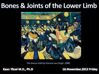
Bones of the lower limb. Krešimir Tućin 2nd year, 2013/14 University of Zagreb Medical School Mentor: A. Žmegač Horvat. Function. C arr y and support the weight of the entire erect body P oints for muscular attachments L ocomotion (standing, walking, jumping, running, kicking, etc.).

E N D
Bones of the lower limb Krešimir Tućin 2nd year, 2013/14 University of Zagreb Medical School Mentor: A. Žmegač Horvat
Function • Carryand support the weight of the entire erect body • Points for muscular attachments • Locomotion (standing, walking, jumping, running, kicking, etc.)
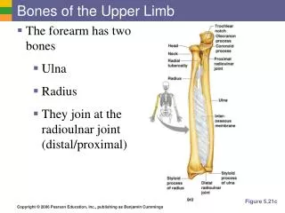
Components • Thigh • Femur • Knee • Patella • Leg • Tibia (medial) • Fibula (lateral) • Foot • Tarsals (7) • Metatarsals (5) • Phalanges (14)
Surface anatomy • Bony prominences and margins – palpable • Exception: hip joint, neck, and shaft of the femur • Anatomical landmarks – define the extent of the leg • Superior anterior iliac spine, greater trochanter, medial condyle of tibia, and medial malleolus
Anterior thigh • Patella • Femur • Condyles • Anterior leg bones • Tibia • Tibialtuberosity • Anterior crest • Medial surface • Medial malleolus • Fibula • Head • Lateral malleolus • Posterior side – only tendons palpable
Femur • Thigh bone • Largest, longest, strongest bone in the body! • Receives a lot of stress • Courses medially • More in women! • Articulates with acetabulum proximally hip (“ball and socket”) joint • Articulates withtibia and patella distally knee joint • lat. acetabulum = vinegar cup
Knee • Femur, patella, tibia • Patella • Kneecap • Triangular sesamoid bone • Protects knee joint • Improves leverage of thigh muscles acting across the knee • Contained within patellar ligament
Tibia • Shin bone • Receives weight of body from femur and transmits to foot • Second to femur in size and weight • Articulates with fibula proximally and distally • Articulates with femur and patella proximally
Fibula • Does NOT bear weight! • Muscle attachment • Not part of knee joint • Stabilizes ankle joint *ankle joint = tibia, fibula, talus
Foot • Function: • Supports the weight of the body • Acts as a lever to propel the body forward • Parts: • Tarsals • Metatarsals • Phalanges of the foot
Tarsals • Hind part of the foot • 7 short bones • Similar to the carpals • Parts: • Calcaneus • Talus • Navicular • 3 Cuneiforms • Cuboid
Metatarsals and phalanges of the foot • Metatarsals • Midfoot • Similar to metacarpals • Phalanges • Forefoot • 2 in the big toe, 3 in each of the other toes • Both long bones
Foot arches • 3 arches • Medial Longitudinal • Lateral • Transverse • Tendons • Inferior to foot bones • Help support arches of foot • Function • Supporttheweightofthebodyduringstanding • Elasticpropulsionduringwalking • Flat foot
References • en.wikipedia.org • www.innerbody.com • www.dartmouth.edu • faculty.ccri.edu • Elsevier, Drake et al: Gray's anatomy for students