Download
1 / 8
80 likes | 144 Views
Explore the intricate anatomy and vital functions of the heart to understand its crucial role in the circulatory system. Dive into the chambers, walls, valves, and vessels of this essential organ.

E N D
Function of the heart • Cells depend on interstitial fluid for survival • The circulatory system balances the contents of the interstitial fluid (gas exchange, nutrients, waste, etc…) • The heart provides the mechanical “pumping” necessary for blood to circulate • Two circuits: • Pulmonary (b/t heart & lungs) • Systemic (b/t heart & body)
Anatomy of the heart • Size: 5” x 3.5” (~fist) • Shape: blunt cone • Apex: pointed end • Base: uppermost part • Location: center of thorax, behind the sternum (mediastinum) • 4 Chambers • Right atrium • Right ventricle • Left atrium • Left ventricle
Pericardial cavity (Fig. 12-2) • Pericardium encloses the heart (fist in balloon) and has 2 layers • Visceral (epicardium): inner layer closest to the surface of the heart • Parietal: outer layer • Pericardial fluid fills the cavity (lubricant)
Surface Anatomy of the heart (Fig. 12-3) • Auricle: outer flap of deflated atrium • Coronary Sulcus: groove between atria and ventricles • Anterior/Posterior Interventricular Sulci: boundary b/t lft. and rt. ventricles
The heart wall (Fig. 12-4) • Three layers • Epicardium: outermost layer (serous) • Myocardium: cardiac muscular layer; concentric wrapping • Endocardium: innermost layer, continuous with vessel linings
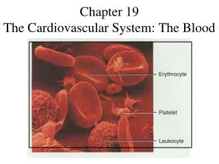
Internal anatomy of the heart • Interatrial Septum: separates left/right atria • Interventricular septum: separates left/right ventricles • Left/Right Atrioventricular (AV) valve • Bicuspid: left AV has 2 cusps, aka mitral valve • Tricuspid right AV has 3 cusps • Superior Vena Cava: blood from head, neck, upper limbs, and chest • Inferior Vena Cava: blood from rest of the trunk, viscera, and lower limbs • Coronary Sinus: opens into right atrium, receives blood from coronary veins
Internal anatomy of the heart • Chordae Tendineae: connects cusps to papillary muscles • Pulmonary semilunar valve: b/t right ventricle and pulmonary arteries • Aortic semilunar valve: b/t left ventricle and aorta • Aorta: start of systemic circuit • Pulmonary trunk: start of pulmonary circuit • Left/Right Pulmonary veins: receives deoxygenated blood from lungs