Download
1 / 52
520 likes | 650 Views
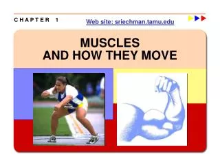
C H A P T E R 1. Web site: sriechman.tamu.edu. MUSCLES AND HOW THEY MOVE. Learning Objectives. w Learn the basic components of skeletal muscle, the muscle fiber, and the myofibril. w Note the cellular events leading to a basic muscle action.

E N D
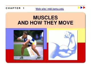
C H A P T E R 1 Web site: sriechman.tamu.edu MUSCLES AND HOW THEY MOVE
Learning Objectives w Learn the basic components of skeletal muscle, the muscle fiber, and the myofibril. w Note the cellular events leading to a basic muscle action. w Discover how muscle functions during exercise. w Consider the differences in fiber types and their impact on physical performance. w Learn how muscles generate force and movement by pulling on bones.
Types of Muscle Cells • Skeletal - striated • w Voluntary muscle; controlled consciously • Over 600 throughout the body Cardiac - striated w Involuntarily controlled (unconscious) by the autonomic nervous system and hormones w Only in the heart • Smooth – non-striated • w Involuntary muscle; controlled unconsciously • In the walls of blood vessels and internal organs
Historical note: striated muscle cells Antony van Leeuwenhoek (1632-1723) developed the microscope, and described the striped appearance of skeletal muscle.
“Sarco” = muscle MUSCLE FIBER Plasma membrane Endoplasmic reticulum (calcium) Cytoplasm “Transmission”-> nerve impulses, waste products
Key Points Muscle Fiber (Multinucleated) w An individual muscle cell is called a muscle fiber or myofiber. w A muscle fiber is enclosed by a plasma membrane called the plasmalemma (sarcolemma). w The cytoplasm of a muscle fiber is called the sarcoplasm. w The t-tubules allow transport of substances and passage of action potentials throughout the muscle fiber. w The sarcoplasmic reticulum stores and releases calcium during contraction.
Key Points Myofibrils wMyofibrils are the contractile “threads” that run the length of the muscle fiber, with several hundred to several thousand within a single fiber. w Myofibrils are made up of sarcomeres in series, the smallest functional units of a muscle. w A sarcomere is composed of filaments of two proteins, myosin and actin, which are responsible for muscle contraction. wThick filament: myosin, which has a globular head at one end (cross-bridge). wThin filament: composed of actin, tropomyosin, and troponin — is attached to the Z disks at the ends of the sarcomeres.
A-bands I-bands H-zone Z-disk Mitochondria Fat M-line Electron Micrograph Showing Myofibrils and Sarcomeres Can you identify the following: 1. sarcomere A-bands, 2. sarcomere I-bands, 3. mitochondria, 4. lipid (fat) droplets, 5. Z-lines, and 6. M-lines ?
Thought Question In the electron micrograph on the preceding slide, can you identify the following: 1. sarcomere A-bands, 2. sarcomere I-bands, 3. mitochondria, 4. lipid (fat) droplets, 5. Z-lines, and 6. M-lines ?
THICK FILAMENT Cross-bridge (actin binding site; ATP binding site; ATPase)
THIN FILAMENT (covers myosinbinding sites at rest) (Ca2+ binding site) (myosin binding site)
5. The Ca2+ binds to troponin on the thin filament, and the troponin pulls tropomyosin off the active sites, allowing myosin heads to attach to the actin filament. (continued) Excitation/Contraction Coupling 1. An α-motoneuron, with signals from the brain or spinal cord, releases the neurotransmitter acetylcholine (ACh) at the neuromuscular junction. 2. ACh crosses the junction and binds to receptors on the plasmalemma (sarcolemma). 3. This initiates an action potential. 4. The action potential travels along the plasmalemma and down the t-tubules to the SR, which releases calcium ions (Ca2+).
Excitation/Contraction Coupling 6. Once a strong binding state is established with actin, the myosin head tilts, pulling the actin filament toward the center of the sarcomere (power stroke). 7. The myosin head binds to ATP, an ATPase found on the head splits ATP into ADP and Pi, releasing energy. ATP ADP + Pi + work + heat myosin ATPase 8. Muscle action continues as long as calcium is elevated, and ends when calcium is actively pumped out of the sarcoplasm back into the sarcoplasmic reticulum by a calcium ATPase.
Thought Question What is the “basic molecule” of exercise physiology? Why?
Exhale CO2 Motor cortex Spinal cord Motor unit Axon Acetylcholine (Neuromuscular junction) Depolarization T-tubules Sarcoplasmic Reticulum CO2 +H20 Inhale O2 2e- + H+ + O2 myoglobin/hemaglobin Electron Transport Chain NADH Food Adipose Krebs Cycle Intramuscular fat β-oxidation Acetyl-CoA Free Fatty Acids Pyruvate Lactate Blood glycolysis Glucose Blood Liver Food Glycogen * * * PCr * Calcium ATP *Note the length of these arrows is related to speed of production (short = fast) and quantity of production capacity (long= large capacity)
MOTOR UNIT CNS (brain, spinal cord)
Sliding Filament Theory w When myosin cross-bridges are activated, they bind strongly with actin, resulting in a conformational change in the cross-bridge. w This change in the cross-bridge causes the myosin head to tilt toward the arm of the cross-bridge and pull the thin filament toward the M-line in the center of the sarcomere. w The tilt of the myosin head is known as a power stroke. w The pulling of the actin filament past the myosin results in muscle shortening and generation of muscle force.
Active State during a Twitch Active state: “true” cross-bridge tension produced Light response: free calcium ion concentration in the sarcoplasm Force: the force measured at the end of the muscle McMahon, Muscles, Reflexes, and Locomotion, 1984
Thought Question In the active state graph on the preceding slide, why is the force measured at the ends of the muscle less than the actual force produced by the cross-bridges?
Muscle Biopsy w Hollow needle is inserted into muscle to take a sample. w Sample is mounted, frozen, thinly sliced, and examined under a microscope. w Allows study of muscle fibers and the effects of acute exercise and exercise training on fiber composition.
Type I (ST) muscle fibers (high concentration in deep antigravity muscles, e.g., soleus) w High aerobic (oxidative) capacity and fatigue resistance 1. High mitochondrial density 2. High capillary density: high blood flow 3. High myoglobin: high oxygen storage and transfer w Low anaerobic (glycolytic) capacity w Relatively slow contractile speed 1. low myosin ATPase activity 2. Relatively little SR (slow Ca2+ release and uptake) w < 300 fibers per motor neuron (relatively small motor units)
Type IIa (FTa) Muscle Fibers w Moderate aerobic (oxidative) capacity and fatigue resistance w High anaerobic (glycolytic) capacity 1. High glycolytic enzymes, thus, high lactic acid production w Fast contractile speed 1. high myosin ATPase activity 2. High density of SR (fast Ca2+ release and uptake) w Relatively large motor units (>300 muscle fibers per motor neuron)
Type IIx (FTb) Muscle Fibers w Low aerobic (oxidative) capacity and low fatigue resistance w High anaerobic (glycolytic) capacity; high lactate production when active w Fast contractile speed (high myosin ATPase and SR density) w Relatively large motor units (>300 muscle fibers per motor neuron)
Strength of the fiber types The higher force development in type II than in type I motor units is due to more muscle fibers per motor unit and larger size of the individual fibers, not the specific force. In other words, a type I and a type II muscle fiber of the same size will produce the same maximal isometric force. Specific force = force/cross-sectional area P. 41
SLOW- AND FAST-TWITCH FIBERS Type I Type IIa Type IIx Acid-stable myosin ATPase
Muscle fiber types w Slow twitch (type I) - #1 below w Fast twitcha (type IIa) - #2 below w Fast twitchb (type IIx(b)) - #3 below Myosin ATPase (Shortening Velocity) Succinate Dehydrogenase Activity (Mitochondrial Density: Oxidative Capacity) Enzyme Histochemistry
Force Production in the Fiber Types The difference in force development between type I and type II motor units is due to the larger number of muscle fibers per motor unit and the larger diameter of the type II fibers.
PEAK POWER GENERATED BY FIBERS Power = force x distance time or Power = work/time or Power = force x velocity For fibers of the same size, force production is about the same in type I and II fibers; however, shortening velocity is much higher in type II fibers
What Determines Fiber Type? w The α-motoneuron of the motor unit controls the properties of the muscle fibers w Demonstrated by: w Cross-innervation of slow and fast muscles McComas, Skeletal Muscle, Human Kinetics, 1996, from Buller et al., 1960.
What Determines Fiber Type? w The α-motoneuron of the motor unit controls the properties of the muscle fibers w Demonstrated by: w Chronic low frequency stimulation McComas, Skeletal Muscle Human Kinetics, 1996, from Salmons and Vrbova, 1969.
What Determines Fiber Type? w Genes determine which motor neurons innervate individual muscle fibers, all fibers in motor units are of the same type. w Both resistance and endurance training and muscular inactivity may result in small changes in the percentages of type I and II fibers. w There is often an increase in the percentage of ST fibers with aging. Skeletal muscles contain both ST and FT motor units; some muscles, like soleus, contain primarily ST; others, like gastrocnemius, are primarily fast-twitch.
The All-Or-None-Response w Fibers in a motor unit are recruited when an alpha-motoneuron carries an action potential (impulse) from the central nervous system. w All muscle fibers in the motor unit are recruited simultaneously. w More force is produced by activating more motor units.
Orderly Recruitment of Muscle Fibers • Principle of orderly recruitment states that motor units are activated in a fixed order • Size principle states that the order of recruitment is directly related to the motor neuron size. • Slow-twitch fibers, which have smaller motor neurons, are recruited before fast-twitch fibers.
Thought Question From a biological perspective, why do you find within a synergistic group of anti-gravity muscles (like the calf muscles) one muscle that is primarily made up of slow fibers (i.e., soleus muscle), and other muscles primarily made up of fast fibers (i.e., gastrocnemius and plantaris muscles)?
Functional Classification of Muscles Agonists—prime movers; responsible for the movement Antagonists—oppose the agonists to prevent overstretching of them Synergists—assist the agonists and sometimes fine-tune the direction of movement
Neural Factors Influencing Force Number of motor units activated (recruitment) Rate of stimulation of the motor unit (rate coding)
Frequency of action potentials (rate coding) Brooks et al., Exercise Physiology, 3rd ed., Mayfield, 2000.
Factors Influencing Force Generation Number of motor units activated Rate of stimulation of the motor unit (rate coding) Type of motor units activated (FT or ST) Muscle size Initial muscle length Joint angle Speed of muscle action (shortening or lengthening)