Download
1 / 5
50 likes | 193 Views
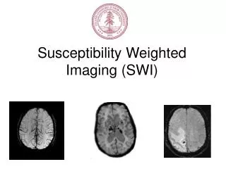
Tumor Imaging and SWI: Possible Directions. WSU MRRC, Imaging Retreat-05. Flash 3D T 1 post-Gad. SWI No-Gad. High Resolution Tumor Imaging-SWI. Surrounding venous vasculature and tumor internal hemorrhage. Also seen are (arrows) micro hemorrhages which are not seen in T 1 weighted images.

E N D
Tumor Imaging and SWI: Possible Directions WSU MRRC, Imaging Retreat-05
Flash 3D T1 post-Gad SWI No-Gad High Resolution Tumor Imaging-SWI Surrounding venous vasculature and tumor internal hemorrhage. Also seen are (arrows) micro hemorrhages which are not seen in T1 weighted images.
High Resolution Tumor Imaging-Flash 3D T1 T1 subtraction maximum intensity projection image Tumor draining/feeding vessels SWI image mIP-8 mm.
Evaluation in Tumor 38 Patients From “Susceptibility Weighted Imaging of Brain Masses” Vivek S et al Submitted to JMRI.
Current Work and Future Goals:Phase filtering and Susceptometry using Fourier based Filter Method