Download
1 / 20
200 likes | 394 Views
Using Iron Porphyrins as Models for Hemoglobin. The system:. Key Features of Hemes Fe oxidation state Fe spin state porphyrin oxidation state porphyrin hydrophobicity. How will the spin state of Fe(porphyrin) complexes change on binding imidazole?. Intermediate Spin S = 3/2

E N D
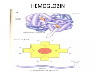
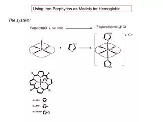
Using Iron Porphyrins as Models for Hemoglobin The system:
Key Features of Hemes • Fe oxidation state • Fe spin state • porphyrin oxidation state • porphyrin • hydrophobicity
How will the spin state of Fe(porphyrin) complexes change on binding imidazole? Intermediate Spin S = 3/2 n = 3 High Spin S = 5/2 n = 5 Low Spin S = 1/2 n = 1
Sample for Evans’ Magnetic Susceptibility Method NMR tube Inside capillary: sample in CHCl3, 1) with imidazole 2) without imidazole Outside capillary: CDCl3 and CHCl3
NMR Spectrum from Evans’ Method Inside capillary: sample in CHCl3, produces broad singlet for paramagnetically shifted CHCl3 below 7.3 ppm Outside capillary: CDCl3 and CHCl3 produces usual sharp singlet for 0.5% CHCl3 at 7.3 ppm
Why is H resonance in CHCl3 shifted downfield and broadened? • pseudocontact and contact terms • addition of new small magnetic field to local magnetic fields • of neighboring nuclei • is used in NMR Shift Reagents to “de-tangle” complicated spectra
Shift of signal, in Hz mass susceptibility of solvent -a diamagnetic contribution, a (-) value Mass susceptibility (+) Magnetic field (400 MHz, or 400 x 106 Hz) Concentration of sample, in g/mL How does shift, , relate to a magnetization of paramagnetic sample? g = 3 0 c
Magnetic field lines of flux Magnetic field lines affected by a paramagnetic substance: attracts Susceptibility, X > 0 Magnetic field lines affected by a diamagnetic substance: repels Susceptibility, X < 0
How does mass susceptibility, g , relate to unpaired electrons in a paramagnetic sample? Mass susceptibility g x (Mol. Wt.) = M Molar susceptibility corr= M - diamagnetic corrections where diamagnetic corrections for Fe, porphyrin, Cl, imidazole, a negative number! eff = 3 R T corr1/2 = 2.828 (T corr) 1/2 N 2 eff = (n(n+2))1/2
Diamagnetic Corrections (cgs units) Xo (CHCl3) = - 4.97 x 10⁻⁷ cgs Porphyrin: TPP= -700 x 10-6 cgs TTP= -753 x 10-6 cgs TClPP= -760 x 10-6 cgs Fe = -13 x 10-6 cgs Cl = -20 x 10-6 cgs Imidazole = -38 x 10-6 cgs
The Role of Axial Ligation and the Allosteric Effect in Hemoglobin O2 Binding
3d orbitals on Fe Spin State of Fe affects size of ion
Large, high spin Fe(2+): In T state, transmitted by His on protein helix Small, low spin Fe(2+): In R state, transmitted to His on protein helix
Use of Cr(acac)3as a Paramagnetic Relaxation Agent in 13C NMR With Cr(acac)3 (note: does not affect chemical shifts) With d1=6.0s (d1: relaxation time)
Use of Paramagnetic NMR in Bioinorganic Systems v m One big mess of piled up H’s on protein!! v m m p p
NMR Paramagnetic Shift Reagents Ground state electron configuration: [Xe] 4f7 6s2 Term Symbol: 8S7/2 how many unpaired e-? EuFOD :also called Eu(fod)3. Eu(OCC(CH3)3CHCOC3F7)3
NMR Paramagnetic Shift Reagents: Eu vs Pr Using Eu(fod)3 oooh! Lovely!! With NO Shift rgt Hmmm, not so pretty Huh? – signals shifted upfield with Pr Using Pr(fod)3
MRI Contrast agents: same principles, applied to medicine • MRI Contrast Agents: observes differential magnetization of protons in different types of molecules that predominate in different tissues. The different magnetization signal intensities produce the contrast between tissues. • The nuclear magnetization is produced by the pulse sequence applied, by the density of nuclear spins sub-fractions (water vs fat protons) and by the spin-lattice relaxation time T1 and phase relaxation time T2 in each nuclear spin sub-fraction. T1 and T2 depend on tissues type. • MRI Contrast Agents interact with one sub-fraction type (usually that easily exchangeable protons, like water) to increase the T1 spin-lattice relaxation times. • The most commonly used compounds for contrast enhancement are gadolinium-based. • MRI contrast agents are used as oral or intravenous administration. Gadoteric acid Effect of contrast agent on images: Defect of the blood–brain barrier after stroke shown in MRI. T1-weighted images, left image without, right image with contrast medium administration.