Download
1 / 39
760 likes | 1.99k Views
Hydrocephalus. From Greek hydrokephalos, from hydr- + kephalE head

E N D
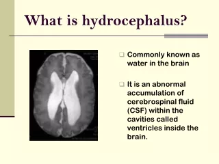
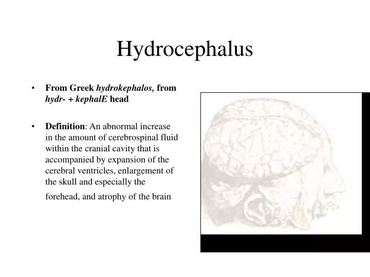
Hydrocephalus • From Greek hydrokephalos, from hydr- + kephalE head • Definition: An abnormal increase in the amount of cerebrospinal fluid within the cranial cavity that is accompanied by expansion of the cerebral ventricles, enlargement of the skull and especially the forehead, and atrophy of the brain
Overview of CSF production • The CSF volume of an average adult ranges from 80 to 160 ml • The ventricular system holds approximately 20 to 50 ml of CSF • CSF is produced in the choroid plexuses at a daily rate of 14-36 ml/hr
Overview of CSF production • The choroid plexuses are the source of approximately 80% of the CSF • The blood vessels in the subependymal regions, and pia also contribute to the formation of CSF
Overview of CSF circulation • The CSF flows from the lateral ventricles downward to the foramina of Magendie and Luschka, to the perimedullary and perispinal subarachnoid spaces, and then upward to the basal cistern and finally to the superior and lateral surfaces of the cerebral hemispheres
CSF circulation • The pressure gradient is highest in the lateral ventricles and diminishes successively along the subarachnoid space • Arterial pulsations in the choroid plexuses help drive the fluid from the ventricular system • Normally, the periventricular tissues offer little resistance to the flow of CSF + -
The CSF volume and pressure are maintained on a minute to minute basis by the systemic circulation CSF pressure is in equilibrium with capillary pressure (determined by the arteriolar tone) An increase in blood PCO2 (hypoventilation) decreases pH and arteriolar resistance, this in turn gives rise to increased CSF pressure by increasing cerebral blood flow Hyperventilation has the opposite effect CSF pressure
CSF pressure • Normal intracranial pressure (ICP) in an adult is between 2-8 mmHg. • Levels up to 16 mmHg are considered normal • ICP higher than 40 mmHg or lower BP may combine to cause ischemic damage
CSF pressure • Increased venous pressure has a direct effect on CSF pressure • Downstream block of venous flow increases the volume of cerebral veins, dural sinuses, and the subarachnoid space
The function of the CSF • The CSF acts as a “water jacket” for the brain and spinal cord • The 1300 g adult brain weighs approximately 45 g when suspended in CSF
The function of the CSF • The CSF acts like a “sink”, effectively flushing waste products as new fluid is secreted reabsorbed • A constant CSF electrolyte composition helps maintain a stable medium for excitable cells (neurons)
Mechanisms of increase intracranial pressure • Brain, Blood and CSF are held in an inelastic container (cranium, vertebral canal and dura) • Changes in volume of either element (Brain,CSF, Blood) is at the expense of the other two
HydrocephalusCommunicating vs. Non-communicating (Dandy) • This is an old classification of hydrocephalus • The terms refer to the presence or absence of a communication of the lateral ventricles with the spinal subarachnoid space
Communicating vs. Non-communicating • This classification was based on the imaging findings after injection of dye into the ventricular system and simultaneous injection of air into the subarachnoid space • Diffusion of dye into the subarachnoid space and passage of air into the ventricular space were the criteria for communicating hydrocephalus
Non-communicating hydrocephalus • There is no communication between the ventricular system and the subarachnoid space. The commonest cause of this category is aqueduct blockage or stenosis.
Aqueductal stenosis • The normal aqueduct measures about 1 mm in diameter, and is about 11 mm in length.
Aqueductal stenosis • Is the most common cause of congenital hydrocephalus(43%) • Aqueduct develops about the 6th week of gestation • M:F = 2:1 • Other congenital anomalies (16%): thumb deformities • Prognosis: 11-30% mortality
Etiology of aqueductal stenosis • Intrinsic Pathology of the Aqueduct • Septum or Membrane Formation: A thin membrane of neuroglia may occlude the aqueduct. It commonly occurs caudally. There may be a primary developmental defect or it may follow granular ependymitis from intrauterine infections. This is the rarest of the types of narrowing. • Forking of the Aqueduct:Typically, there are two channels seen in midsagittal plane unable to handle CSF volume. Most often seen with spina bifida. • Gliosis of the Aqueduct: Usually of infectious origin showing a marked gliofibrillary response. The lumen is devoid of ependyma. • Stenosis of the Aqueduct: Narrowed aqueduct without evidence of gliosis. This may have hereditary basis.
Etiology of aqueductal stenosis • Extrinsic Pathology of the Aqueduct: • Infectious. Abscesses. • Neoplastic. Pineal tumors, brainstem gliomas, medulloblastoma, ependymoma. • Vascular. AVM, aneurysm, Galen aneurysm. • Developmental. Arachnoid cysts.
Clinical features of aqueductal stenosis • Obstructive hydrocephalus: presents with macrocephaly and/or intracranial hypertension. • Parinaud's syndrome. Inability to elevate eyes • Collier's sign. Retraction of the eyelids
Imaging of aqueductal stenosis • Ultrasonography can detect aqueductal stenosis in utero. Sonogram
Imaging of aqueductal stenosis • CT and MRI. MRI is essential if third ventriculostomy is to be considered.
Treatment of aqueductal stenosis • Treatment and Results • Remove underlying cause of obstruction if possible. • Third ventriculostomy as initial treatment of choice. • VP shunt if technical reasons do not allow third ventriculostomy or if the child fails after ventriculostomy. • Aqueductal stent can be placed if technically feasible. Usually rarely done due to risk of upper brain stem injury.
Communicating hydrocephalus • In communicating or non-obstructive hydrocephalus there is communication between the ventricular system and the subarachnoid space. The commonest cause of this group is post-infectious and post-hemorrhagic hydrocephalus.
Causes of communicating hydrocephalus • Overproduction of CSF • Blockage of CSF circulation • Blockage of CSF resorption • Hydrocephalus ex-vacuo • Normal pressure hydrocephalus
Overproduction of CSF • Excessive secretion of CSF by the choroid plexus as in cases of choroid plexus papilloma or carcinoma. This is a rare cause.
Blockage of CSF circulation • This could be at any level of the CSF circulation. It could be at the level of the foramen of Monro, with either unilateral or bilateral occlusion of the foramen of Monro giving dilatation of one or both lateral ventricles. This is commonly seen in the colloid cyst and tumors of the third ventricle.
Dandy Walker Syndrome • A common cause of obstructive hydrocephalus is Dandy Walker Syndrome where there is blockage of foramina of the 4th ventricle. This is a congenital condition associated with agenesis of the cerebellar vermis
Blockage of CSF resorption • Poor resorption of CSF into the venous sinuses caused by scarring of the arachnoid villi and is commonly seen after meningitis or hemorrhage
Hydrocephalus Ex Vacuo • Hydrocephalus ex-vacuo involves the presence of too much CSF, although the CSF pressure itself is normal. This condition occurs when there is damage to the brain caused by stroke or other form of injury or chronic neurodegeneration, and there may be an actual shrinkage of brain substance.
Normal pressure hydrocephalus • Normal pressure hydrocephalus (NPH) is usually due to a gradual blockage of the CSF drainage pathways in the brain. NPH is an unusual cause of dementia, which can occur as a complication of brain infection or bleeding (hemorrhage).
In some patients, no predisposing cause can be identified. In patients with NPH, although the ventricles enlarge, the pressure of the CSF remains within normal range. NPH is characterized by gradual memory loss (dementia), balance disorder (ataxia), urine incontinence, and a general slowing of activity. Symptoms progressively worsen over weeks. In some patients, an improvement of symptoms is noted immediately after the removal of spinal fluid with a lumbar procedure. Normal pressure hydrocephalus
Treatment of hydrocephalus • The two most commonly used shunt systems are the ventriculoatrial (VA) and ventriculoperitoneal (VP) shunts. The VP shunt is most commonly used as it is simpler to place, extra tubing may be placed in the peritoneum and the consequences of infection are less.
Treatment of hydrocephalus • The VA shunt must be accurately located in the atrium and requires frequent revisions as the child grows to maintain the proper position of the distal end. In addition, infection is a more serious complication with a VA shunt as its location in the blood stream may lead to sepsis.
Treatment of hydrocephalus • Recently, in situations where both the abdomen and vascular system can no longer function to absorb CSF, Pediatric Neurosurgeons have begun to place the distal catheter in the pleural space (V-PL shunt). The distal catheter is placed through a small incision in the anterior chest wall. As with the peritoneal shunt, extra tubing can be placed, reducing the need for further shunt revisions.
Treatment of hydrocephalus • Shunt systems include three components: (1) a ventricular catheter, (2) a one way valve and (3) a distal catheter. The ventricular catheter is a straight piece of tubing, closed on the proximal end and usually with multiple holes for the entry of CSF along the proximal two centimeters of the tube.
Treatment of hydrocephalus • Shunts are composed of a material called Silastic. Silastic is made from a family of polymerized organic compounds termed silicone. Silicone is the substance that has caused controversy in breast implants because of the association with auto immune disorders. So far no cases of auto immune disease have been linked to the Silastic used in shunts.
Treatment of hydrocephalus • The most common sites for entry of the ventricular catheter are a frontal position in line with the pupil at the coronal suture, a parietal position just above and behind the ear, or an occipital position three centimeters off the posterior midline. The position used varies with the configuration of the ventricles, the shape and size of the head and the surgeon’s preference.
Shunt malfunction • Common complications of VP shunt include shunt malfunction or blockage and infection. Malfunction may be related to growth and the shunt will need to be replaced with a longer catheter. Symptoms of shunt malfunction or infection include headache, fever, drowsiness,convulsions, increased head circumference and bulging fontanelle.
Shunt malfunction • If left untreated, shunt malfunction or infection is associated with high morbidity and mortality rates. Most patients with these complications have subtle presentations and nonspecific signs, despite elevated ICP or CNS infection. The workup includes a focused review of records, information from the patient’s family or caretaker, and elements of a unique examination to supplement routine work-up of the patient with a ventricular shunt. A shunt series and head CT scan are part of the initial evaluation. Empiric antibiotic therapy is initiated to cover Gram-positive organisms, predominantly S. epidermidis, as well as the less common Gram-negative and anaerobic organisms responsible for shunt infections.