Download
1 / 24
260 likes | 423 Views
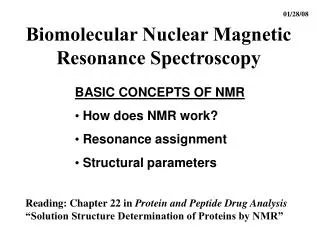
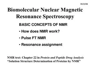
01/22/03. Biomolecular Nuclear Magnetic Resonance Spectroscopy. BIOCHEMISTRY BEYOND STRUCTURE Protein dynamics from NMR Analytical Biochemistry Comparative Analysis. Why The Interest In Dynamics?. Function requires motion/kinetic energy Entropic contributions to binding events

E N D
01/22/03 Biomolecular Nuclear Magnetic Resonance Spectroscopy BIOCHEMISTRY BEYOND STRUCTURE • Protein dynamics from NMR • Analytical Biochemistry • Comparative Analysis
Why The Interest In Dynamics? • Function requires motion/kinetic energy • Entropic contributions to binding events • Protein Folding/Unfolding • Uncertainty in NMR and crystal structures • Effect on NMR experiments-spin relaxation is dependent on rate of motions know dynamics to predict outcomes and design new experiments • Quantum mechanics/prediction (masochism)
Dynamics From NMR Parameters • Number of signals per atom: multiple signals for slow exchange between conformational states Populations ~ relative stability Rex < w (A) - w (B) Rate A B
Dynamics From NMR Parameters • Number of signals per atom: multiple signals for slow exchange between conformational states • Linewidths: narrow = faster motion, wide = slower; dependent on MW and structure
B A B A 15N 15N 15N 1H 1H 1H Linewidth is Dependent on MW • Same shifts, same structure • Linewidth determined by size of particle • Fragments have narrower linewidths
40 173 P Detecting Functionally Independent Domains in Multi-Domain Proteins RPA32 RPA14 Why? • Flexibility facilitates interactions with protein targets
Dynamics From NMR Parameters • Number of signals per atom: multiple signals for slow exchange between conformational states • Linewidths: narrow = faster motion, wide = slower; dependent on MW and conformational states • Exchange of NH with solvent:slow timescales (milliseconds to years!) • Requires local and/or global unfolding events • NH involved in H-bond exchanges slowly • Surface or flexible region: NH exchanges rapidly
Dynamics From NMR Parameters • Number of signals per atom: multiple signals for slow exchange between conformational states • Linewidths: narrow = faster motion, wide = slower; dependent on MW and conformational states • Exchange of NH with solvent:slow timescales • NMR relaxation measurements (ps-ns, ms-ms) • R1 (1/T1) spin-lattice relaxation rate (z-axis) • R2 (1/T2) spin-spin relaxation rate (xy-plane) • Heteronuclear NOE (e.g. 15N- 1H)
Dynamics To Probe The OriginOf Structural Uncertainty Weak correlation • Measurements show if high RMSD is due to high flexibility (low S2) Strong correlation
Analytical Protein Biochemistry • Purity (1-2%)- heterogeneity, degradation, buffer • Check on sequence (fingerprint regions) • Binding constants, off rates, on rates
Protein Fingerprints 1H COSY 15N-1H HSQC 13C HSQC also! Assay structure from residue counts in each fingerprint
Monitoring Binding Events Titration followed by 15N-1H HSQC NMR Provides • Site-specific • Multiple probes • In-depth information • Spatial distribution of responses can be mapped on structure
Binding Constants From NMR Stronger Weaker Molar ratio of d-CTTCA Fit change in chemical shift to binding equation
Comparative Analysis • Different preparations, chemical modifications • Conformational heterogeneity (e.g. cis-trans isomerization) • Homologous proteins, mutants, engineered proteins
B A B A Comparative Analysis of StructureIs the protein still the same when we cut it in half? RPA70 15N If the peaks are in the same place, the structure is the same 15N 15N 2 2 3 1H 3 1 1 1H 1H Same idea for comparing mutants or homologs
Biochemical Assay of MutationsMutations can effect folding and stability Wild-type Partially destabilized Partially destabilized & hetero-geneous Unfolded
Biochemical Assay of MutationsWhat is the cause of the Prp19-1 defect? Not perturbation at binding interface Destabilized U-box leads to drop in activity
C N Winged Helix-Loop-Helix Probing Binding of Protein TargetsStructure is the Starting Point! Mer et al., Cell (2000)
Probe Binding Events by NMR15N-RPA32C + Unlabeled XPA1-98 15N-1H HSQC • Only 19 residues affected • Discrete binding site • Signal broadening exchange between the bound and un-bound state • Kd > 1 mM RPA32C RPA32C + XPA 1-98
C N NMR Identification of theXPA Binding Site on RPA32C Map of chemical shift perturbations on the structure of RPA32C
Localization of Binding Site • Same residues bind to peptide and protein • Same binding site • Slower exchange for peptide • Kd < 1 mM XPA1-98 domain XPA29-46 peptide
Manual Database Search Predicts Binding Sites in Other DNA Repair Proteins XPA29-46 UDG79-88 RAD257-274 E R K RQR A L ML R QA R L A A R R I Q RNK A A AL L RL A A R R K L RQK Q L Q Q Q F R E R M E K
All Three Proteins Bind to RPA32CBinding Sites are Identical UDG79-88 RAD257-274 XPA29-46