Download
1 / 39
390 likes | 793 Views
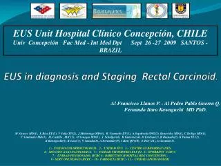
EUS Unit Hospital Clínico Concepción, CHILE Univ Concepción Fac Med - Int Med Dpt Sept 26 -27 2009 SANTOS - BRAZIL . EUS in diagnosis and Staging Rectal Carcinoid . Al Francisco Llanos P. - Al Pedro Pablo Guerra Q.

E N D
EUS Unit Hospital Clínico Concepción, CHILE Univ Concepción Fac Med - Int Med Dpt Sept 26 -27 2009 SANTOS - BRAZIL EUS in diagnosis and Staging Rectal Carcinoid. Al Francisco Llanos P. - Al Pedro Pablo Guerra Q. Fernando Itaro Kawaguchi MD PhD. M. Ocares MD(1), L Rios EU(3), V Salas TP(2), J Madariaga MD(4), K. Caamaño EU(3), A Sepulveda ING(3), Benavides MD(1), C Zuñiga MD(1), C Santander MD(1), JL Castillo , MsC(5), O Venegas MD(5), J Schalper(4), R Gutierrez(4), D Esteban(2), R Branada(2), K Palma EU(2), K Kawaguchi(6), R Luco(7), V Sawada(8), A Fernandez(9), I Ruiz QF(10), E Eriz (11), A Guzman(3). 1.- UNIDAD COLOPROCTOLOGIA. 2.- UNIDAD EUS 3.- CENTRO GI BIOARRAYANES, 4.- SECCION ANAT.PATOLOGICA. 5.- UNIDAD CITOMETRIA FLUJO. 6.- OTORRINO U DE C. 7.- UNIDAD PENSIONADO, HCRC 8.- DIRECCION HOSPITAL REG CONCEPCION.- 9.- SERV ONCOLOGIA HCRC.- 10.- FARMACIA HCRC.- 11.- UNIDAD APOYO DOLOR.
Colo-rectal Cancer • Increasing incidence(> 900 patients per year in last decade). • Gastrointestinal cancer is prevalent in Chile. • GI cancer is the third cause of death in our country: males and females. • In males, rectal cancer is the most frequent colo-rectal cancer. • Encouraging prognosis. • Found in 8 per 100,000 post-mortem analysis. R. Avendaño, P. Fernández, M. Deichler, “Poliposis de colon” published in “Cuadernos de cirugía v.21 n.1” 2007, Universidad Austral de Chile.
Distribution of 13,715 NETs I.Modlin et al 2007 Am J Gastro
CARCINOID INCIDENCE All sites Digestive system Modlin, SEER 9 Registry (1973-2002)
Incidence – 2003 GI Carcinoids and Adenocarcinomas Adenocarcinomas Carcinoids
White males 1992-1999 (1969-1971) White females 1992-1999 (1969-1971) Black males 1992-1999 (1969-1971) Black females 1992-1999 (1969-1971) 3.98 (1.87) 2.47 (1.31) 2.58 (1.63) 4.48 (2.16) Carcinoid Tumors • SEER data (Surveillance Research Programme of NCI) 1973-1999: 13715 cases Modlin et al Cancer 2003 Incidence per 100,000 population: Third National Cancer Survey 1969-71 and SEER 1992-99 ALS: Francisco Llanos Prado - Pedro Guerra Quintana - Fernando Kawaguchi P. MD PhD
Carcinoid Tumor: • USA: The age-adjusted incidence has increased in the last 35 years. • 55% Gastrointestinal location. • 30% Broncho pulmonary location. • Gut tumors 13 to 34% are carcinod tumors. • Inside GI tract (SEER database): • 45% Distal small intestine, mainly: ileum. • 20% Rectum. • 16% Vermiform appendix. • 11% Colon. • 7% Stomach. ALS: Francisco Llanos Prado - Pedro Guerra Quintana - Fernando Kawaguchi P. MD PhD
Rectal CarcinoidSummary • Arises from etherochromaffin cells. • NET 5HIAA urinary test. • No sexual oragerelatedsusceptibility. • Over-represented among the black and • Asian populations. ALS: Francisco Llanos Prado - Pedro Guerra Quintana - Fernando Kawaguchi P. MD PhD
Rectal Carcinoid • Mainlyasymptomatic, however , decreases fecal quantity, causes rectorragy, pain and rectal tenesmus. • Whensicknessbecomessymptomatic(Carcinoid sindrome) metastasesbecome more frequent: ALS: Francisco Llanos Prado - Pedro Guerra Quintana - Fernando Kawaguchi P. MD PhD
Rectal Carcinoid • Staging T and Metastases: • Lessthan 1 cm, metastases are infrequent. • Among 1 - 1.9 cm, 10% metastases • Largerthan 2 cm, 70% metastases • Anothercriterion: -Muscularispropriainvasion. • -Lympho-vascular invasion. • -High mitotic rate. ALS: Francisco Llanos Prado - Pedro Guerra Quintana - Fernando Kawaguchi P. MD PhD
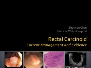
Rectal Carcinoid ALS: Francisco Llanos Prado - Pedro Guerra Quintana - Fernando Kawaguchi P. MD PhD
Rectal Carcinoid ALS: Francisco Llanos Prado - Pedro Guerra Quintana - Fernando Kawaguchi P. MD PhD
Rectal Carcinoid Differents classifications Typical: Uniform cell patterns, rare mitotic activity, no evidence of local invasive growth, bloodstream, lymphatic vessels or neuronal structures. Local invasion or cell pleomorphism. Atypical: WHO clasification(NETs): Well-differentiated : -Benign Behaviour: -Uncertain Behaviour -Low Grade Malignant Behaviour: Poorly differentiated -High Grade Malignant Potential NET = benign/uncertain behavior NEC = some degree of malignancy ALS: Francisco Llanos Prado - Pedro Guerra Quintana - Fernando Kawaguchi P. MD PhD
Rectal Carcinoid Staging &Prognosis SEER database 1977-2004 pts:4704, Landry MD, ELSEVIER GLOBAL MEDICAL NEWS ALS: Francisco Llanos Prado - Pedro Guerra Quintana - Fernando Kawaguchi P. MD PhD
Francisco Llanos Prado - Pedro Guerra Quintana - Fernando Kawaguchi P.
Francisco Llanos Prado - Pedro Guerra Quintana - Fernando Kawaguchi P.
Rectal Carcinoid “Many carcinoid tumors are discovered incidentally during endoscopy or radiographic procedures planned for other purposes” ALS: Francisco Llanos Prado - Pedro Guerra Quintana - Fernando Kawaguchi P. MD PhD
“Many carcinoid tumors are discovered incidentally during endoscopic or radiographic procedures planned for other purposes” AMERICAN GASTROENTEROLOGICAL ASSOCIATION GUIDELINES: “Endoscopy alone is not reliable for detecting the etiology of a subepithelial gastric mass. ” “Cross-sectional imaging techniques such as transabdominalultrasonography, computed tomography, and magnetic resonance imaging are adequate for detecting the presence of normal or abnormal structures outside the gastric wall but do not reliably distinguish between the various causes of masses arising within the gastric wall ” “Endosonographic characterization of subepithelial lesions of the upper gastrointestinal tract” published in www.uptudate.com february 11, 2009. Mary Lee Krinsky, DO & Kenneth Binmoeller, MD” ALS: Francisco Llanos Prado - Pedro Guerra Quintana - Fernando Kawaguchi P. MD PhD
Traditionally, the diagnosis of rectal carcinoid tumors mostly depends on digital examinations, barium enema and colonoscopy. Using these procedures, it is difficult to determine the real size, layer of the origin, and histological nature of the tumor. Zhou PH, Yao LQ, He GJ, Gao WD, Qin XY. Clinical. application of ultrasonic probing for preoperative staging of colorectal carcinoma. Asia J Surg 2003; 26: 18-20. Federspiel BH, Burke AP, Sobin LH, Shekitka KM. Rectal and colonic carcinoids: a clinicopathologic study of 84 cases. Cancer 1990; 65: 135-140. ALS: Francisco Llanos Prado - Pedro Guerra Quintana - Fernando Kawaguchi P. MD PhD
Ultrasound Endoscopy • It is a medical procedure which combines endoscopy and ultrasound to obtain images of internal organs in chest and abdomen • It can be used to visualize the wall of these organs, and adjacent structures • Combined with Doppler imaging, nearby blood vessels can also be evaluated. • It allows biopsies and associate with FNA . • Approximate 10 - 15 minutes . • Allows intraoperative monitoring and follow up. • Allows endoscopic excision by itself. ALS: Francisco Llanos Prado - Pedro Guerra Quintana - Fernando Kawaguchi P. MD PhD
Ultrasound Endoscopy • It provides an understanding of whether the lesion arises from the bowel wall (intramural) or from a structure outside the bowel wall (extramural) compressing the gastrointestinal wall. • It can determine the originating layer of intramural lesions, an important clue for achieving a diagnosis. • The echogenicity, vascularity, margins, size of the lesion, and absence or presence of adjacent lymphonodes also helps to narrow the differential diagnosis. • Provides a minimaly invasive treatment for these tumors. ALS: Francisco Llanos Prado - Pedro Guerra Quintana - Fernando Kawaguchi P. MD PhD
AMERICAN GASTROENTEROLOGICAL ASSOCIATION GUIDELINES: “EUS isthemostaccurateimaging test fordetectingthecomponent of thegastricwallfromwhichthemassarises, informationwhich, combinedwiththeechogenicity of themass, helpsnarrowthedifferential diagnosis ” “Endosonographic characterization of subepithelial lesions of the upper gastrointestinal tract” published in www.uptudate.com february 11, 2009. Mary Lee Krinsky, DO & Kenneth Binmoeller, MD” ALS: Francisco Llanos Prado - Pedro Guerra Quintana - Fernando Kawaguchi P. MD PhD
ALS: Francisco Llanos Prado - Pedro Guerra Quintana - Fernando Kawaguchi P. MD PhD
Accuracy in staging T “TEUS correctly identified 62 percent of patients with T3/4 disease that were missed by CT” Sensitivity and specificity TEUS Endoscopic ultrasound in rectal cancer Gavin C Harewood, MD & Mauritis J Wiersema, MD, may 2009 ALS: Francisco Llanos Prado - Pedro Guerra Quintana - Fernando Kawaguchi P. MD PhD
“TheEndosonographyexceedsthecapacity of otherteststoidentifythedegree of depth of transmuralinvasion and thepresence of peri-rectal nodesinvolved in rectal cancer.” EUS FNA Biopsy “Clinical manifestations, diagnosis, and staging of colorectal cancer” Published www.uptudate.com april 17, 2009. Dennis J Ahnen, MD & Finlay A Macrae, MD ALS: Francisco Llanos Prado - Pedro Guerra Quintana - Fernando Kawaguchi P. MD PhD
EUS together FNA increases even more the accuracy in staging (N) “Clinical manifestations, diagnosis, and staging of colorectal cancer” Published in www.uptudate.com en abril 17, 2009. Dennis J Ahnen, MD & Finlay A Macrae, MD ALS: Francisco Llanos Prado - Pedro Guerra Quintana - Fernando Kawaguchi P. MD PhD
Comparative cost-time requests ALS: Francisco Llanos Prado - Pedro Guerra Quintana - Fernando Kawaguchi P. MD PhD
• According to the guidelines for treating rectal carcinoid tumors, less • than 10 mm with SM involvement have a low risk of metastases, and • endoscopic excision is considered curative. • • As for the lesions measured between 10 mm and 15 mm with SM • involvement, local excision and regional lymphadenectomy is • desirable. • • As a minimally invasive technique, endoscopic resection offers the promise of localized treatment of these tumors, with relatively few complications and rare mortality. • • Various endoscopic Rectal Carcinoid resection, such as endoscopic polypectomy, strip biopsy, aspiration resection, band-snare resection have been described as effective treatments Francisco Llanos Prado - Pedro Guerra Quintana - Fernando Kawaguchi P.
EUS RECTAL CARCINOID RESECTION • However, complete resection of carcinoid tumors of the rectum is difficult with conventional polypectomy, because nearly 75% of the tumors extend into the submucosa. • Fujimura Y, et al. A carcinoid tumor of the rectum removed by strip biopsy. Endoscopy 1993; 25: 428-430. • 8. Kobayashi K, et al. Indications of endoscopic polypectomy for rectal carcinoid tumors and clinical usefulness of endoscopic ultrasonography. Dis Colon Rectum 2005; 48: 285-291. • 9. Jung IS, Ryu CB, Kim JO, . Rectal carcinoid treated by EMR. GastrointestEndosc 2003; 58: 253-255. • 10. Matsumoto T. EUS in rectal carcinoidtumors: contribution to selection of therapy. GastrointestEndosc 1991; 37: 539-542. ALS: Francisco Llanos Prado - Pedro Guerra Quintana - Fernando Kawaguchi P. MD PhD
ALS: Francisco Llanos Prado - Pedro Guerra Quintana - Fernando Kawaguchi P. MD PhD
“This is the most accurate scan to assess tumor size, and ideal for showing the indemnity of the muscularispropria, being these two factors seem to conditionate the possibility of distant metastases. Thus, we will be able to direct accurate therapeutic strategies, in both presurgical management, and neoadjuvant therapy as also postsurgical monitoring. In other hand offers the promise of localized treatment for these tumors.” ALS: Francisco Llanos Prado - Pedro Guerra Quintana - Fernando Kawaguchi P. MD PhD
Chromogranin A • Plasma chromogranin A (CGA) is a more sensitive marker than urinary 5-HIAA in patients with: • -carcinoid tumors • -functioning pancreatic islet cell tumors • -nonfunctioning pancreatic islet cell tumors • -higher level; in patients with diffuse metastases • Plasma CGA levels also have prognostic value • Urinary 5-HIAA • Serial measurement of 5-HIAA in 24-hour urine for carcinoid tumors • Elevated urinary 5-HIAA levels are highly specific for carcinoid tumors • 5-HIAA levels are most useful with primary midgut carcinoid tumors • Foregut and hindgut carcinoids only rarely secrete serotonin • PATHOLOGY • CLASSIFICATION SYSTEMS NOT UNIFORM • NO VALIDATED PROGNOSTIC SYSTEM • STAINING, EXTENT, Ki 67, MITOTIC COUNT, HORMONE PRODUCT • PAUCITY OF PATHOLOGICAL AWARENESS
EUS SM SIMPLE MINIMALLY INVASIVE TREATMENT • SM EUS lesions requires special treatment for deeper resection to achieve clear margins. • This can be done by lifting the lesion, either with SM injection of saline solution (conventional EMR) or by aspiration and banding (ESMR), followed by snare resection. • Kajiyama T, et al. Endoscopic resection of gastrointestinal submucosal lesions: a comparison between strip biopsy and aspiration lumpectomy. GastrointestEndosc 1996; 44: 404-410. • Oshitani N, et al. Endoscopic resection of small rectal carcinoid tumors using an aspiration method with a transparent overcap. J Int Med Res 2000; 28: 241-246. • Nagai T, et al. Saline-assisted endoscopic resection of rectal carcinoids: cap aspiration method versus simple snare resection. Endoscopy 2004; 36: 202-205. • Ono A, et al. Endoscopic submucosal resection of rectal carcinoid tumors with a ligation device. GastrointestEndosc 2003; 57: 583-587. Francisco Llanos Prado - Pedro Guerra Quintana - Fernando Kawaguchi P.
EUS SM SIMPLE MINIMALLY IMVASIVE TREATMENT • Placing the snare below the band, ESMR provides a significant deeper vertical resection margin and, theoretically, a higher rate of curative resection. • In a word, EUS offers new possibilities for the diagnosis of rectal carcinoid tumors. • ESMR is a simple, minimally invasive, and safe procedure for rectal carcinoidtumors. • Kajiyama T, et al. Endoscopicresection of gastrointestinal submucosal lesions: a comparison between strip biopsy and aspiration lumpectomy. GastrointestEndosc 1996; 44: 404-410. • Oshitani N, et al. Endoscopic resection of small rectal carcinoid tumors using an aspiration method with a transparent overcap. J Int Med Res 2000; 28: 241-246. • Nagai T, et al. Saline-assisted endoscopic resection of rectal carcinoids: capaspirationmethod versus simple snareresection. Endoscopy 2004; 36: 202-205. • Ono A, et al. Endoscopic submucosal resection of rectal carcinoid tumors with a ligationdevice. GastrointestEndosc 2003; 57: 583-587.
Tissue markers with diagnostic and prognostic use in GEP-NET Modlin et al. Lancet Oncology 2008; 9(1):61-72
EUS Unit Hospital Clínico Concepción, CHILE Univ Concepción Fac Med - Int Med Dpt Sept 26 -27 2009 SANTOS - BRAZIL EUS in diagnosis and Staging Rectal Carcinoid. M. Ocares MD(1), L Rios EU(3), V Salas TP(2), J Madariaga MD(4), A Heisser EU(3), A Sepulveda ING(3), Benavides MD(1), C Zuñiga MD(1), C Santander MD(1), JL Castillo , MsC(5), O Venegas MD(5), J Schalper(4), R Gutierrez(4), D Esteban(2), R Branada(2), K Umaña EU(2), K Kawaguchi(6), R Luco(7), V Sawada(8), A Fernandez(9), I Ruiz QF(10), E Eriz (11), A Guzman(3). 1.- UNIDAD COLOPROCTOLOGIA. 2.- UNIDAD EUS 3.- CENTRO GI BIOARRAYANES, 4.- SECCION ANAT.PATOLOGICA. 5.- UNIDAD CITOMETRIA FLUJO. 6.- OTORRINO U DE C. 7.- UNIDAD PENSIONADO, HCRC 8.- DIRECCION HOSPITAL REG CONCEPCION.- 9.- SERV ONCOLOGIA HCRC.- 10.- FARMACIA HCRC.- 11.- UNIDAD APOYO DOLOR. ALS: Francisco Llanos Prado - Pedro Guerra Quintana - Fernando Kawaguchi P. MD PhD