Download
1 / 42
420 likes | 529 Views
Explore the layers of skin and dynamics of blood flow in the human body during exercises. Learn how blood flow distribution changes during physical activities and the role of skin in thermoregulation processes.

E N D
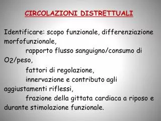
CIRCOLAZI O NE VALORI B A - SCOPO PARTIC O - ADATTAME N - ADATTAMENTI METODI DI SALI LARE TI STRUTT U - FUN ZIONALI M I SURA ml/min/100 g RALI 70-80 portare O al cuore densità capil l - alta estraz. O termodiluiz. 2 2; 300-400 senza interruzione lari; seno coron.; iperemia met a - angiogr a fia bolica MUSCOLO 3-5 15 irrorare un tessuto fibre fasiche e elevato tono va- pletismografia; SCHELETRICO > 60 in cui il metabolismo fibre toniche somotore; iper e - Doppler cambia molto; reg o - mia reattiva; lare RPT debito di O 2 10 -20 anastomosi A- risposta alla 1 - 200 V; disposizione tem peratura l o - controco r rente cale; controllo ipotala mico 55 assicurare flusso di circolo di Wi l - autoregolazione; traccianti r a - 100 (m. gr i - O per un consumo lis; arterie risposta di dioat tivi; Do p - 2 gia) costan temente el e - lunghe Cushing pler caro tideo vato CORONARICA CUTANEA termoregolazione termografia CEREBRALE
This diagram shows the layers found in skin. There are three main layers: the epidermis, dermis and hypodermis. There are also sweat glands, and hairs, which have sebaceous glands, and a smooth muscle called the erector pili muscle, associated with them. Hairs are only found in thin skin, and not in the thick skin present on the fingertips, palms and soles of your feet. Find out more about hair.
Figure 1. Central hemodynamics during incremental exercise to exhaustion. Left column: cardiac output, heart rate (HR), stroke volume, mean arterial (•) and central venous (▾) pressures, and systemic vascular conductance. Right column: arterial O2 content, systemic O2 delivery, systemic a‐vO2 difference, systemic O2 extraction, and systemic O2 uptake during incremental exercise to exhaustion plotted against the relative increase in power output. Data are means ± SEM for eight subjects. * Lower than 80% of peak power, P < 0.05. Adapted from (951) with permission of the Physiological Society and Wiley‐Blackwell
Figure 2. Regional distribution of cardiac output during various intensities of exercise expressed as % maximal oxygen consumption. Bar graphs on the left illustrate total cardiac output (sum of all tissue flows) and the regional distribution of cardiac output to all tissues with increasing exercise intensity. Bar graphs on the right show an expansion of regional blood flow (BF) data to all tissues except skeletal muscle tissue, to enhance appreciation of the changes in BF to the other tissue with exercise (Note the change in the BF axis for the graphs on the right). Data are estimated for human values using Rowell's text book (1172) (also estimates for humans) and our data for regional BF in pigs (29, 30, 239).
Figure 8. Distribution of total CO among legs, respiratory muscles (RM), and other metabolically active tissues (skin, heart, brain, kidneys, and liver) during exercise at o2 max. Adapted from (489) with permission of the American Physiological Society.
Figure 10. Arm blood flow (ABF) and a‐vO2 difference during arm cranking to exhaustion in rowers and average, fit subjects. A + L, addition of leg exercise to arm cranking in the rowers. Values are means ± SE for eight average fit subjects and seven rowers. Adapted from Volianitis et al. (1429) with permission of the American Physiological Society.
Figure 31. Schematic description of the thermoregulatory control of skin blood flow (BF) as modified by moderately intense exercise. The relation of skin BF to internal temperature is affected, relative to resting conditions, in at least three ways by exercise: a vasoconstrictor response at the onset of dynamic exercise (A), an increase in the internal temperature threshold at which skin BF begins to increase (B), and a leveling off, or plateau, in skin BF above an internal temperature of 38°C at a level well below maximal (C). Exercise exerts these effects through the vasoconstrictor system for the initial vasoconstriction and through inhibiting the active vasodilator system for the increased threshold and for the plateau. At rest, the plateau only occurs as skin BF approaches maximal vasodilation. Modified from Gonzalez‐Alonso et al. 2008 (419) with permission of the Physiological Society and Wiley‐Blackwell
Figure 33. Relationship between splanchnic blood flow (BF) and exercise intensity (expressed as percent o2 max) at normal temperature (25.6°C, solid line) and during heat stress (43.3°C, dashed line). The reduction of splanchnic BF during exercise is proportional to the relative exercise intensity (%o2 max) and is further reduced by environmental heat stress resulting in ∼20% greater reductions in splanchnic BF at a given relative exercise intensity. Modified from Rowell, 1993 (1177) which was adapted from Rowell 1965 (1180) with permission of the American Physiological Society.
Figure 34. Exercise‐induced reductions in both splanchnic blood flow (BF) and splanchnic blood volume are nonuniformly distributed across the splanchnic organs. (A) Nonuniform changes in splanchnic BF during and 2 minutes after termination of cycling. The decreases in total splanchnic BF (SBF) (top panel) during moderate cycling exercise involve greater reductions in celiac artery BF (dotted line; middle panel) than in the superior mesenteric artery BF (solid line; middle panel) owing to differential increases in artery resistance (R) in the celiac (dotted line) and superior mesenteric artery (solid line) vascular beds (bottom panel). For both BF and R data in the middle and bottom panels, respectively; continuous line = superior mesenteric artery and the dashed line = the celiac artery. * = different from the preceding value, P < 0.05; # = different from rest, P < 0.05. Data from Perko et al. (1079) with permission of the American Physiological Society. (B) Nonuniform changes in splanchnic blood volume. Splanchnic blood volume redistribution during zero‐load (ZLC) and graded cycling exercise (50%‐100% maximal exercise) and 5 min of recovery from exercise. Note that the volume redistribution involves a greater reduction of splenic than hepatic and renal blood volume and that bowel blood volume did not significantly change from baseline values during or following exercise. Values are % basal volume, mean ± SE.
Figure 35. Exercise training alters the redistribution of blood flow (BF) to kidney and splanchnic tissues during exercise. Top panel: baseline/resting BF values represent sedentary rats standing quietly on the treadmill. Middle panel: exercise BF values for sedentary rats at 15 min of treadmill running 30 m/min. Bottom panel: exercise BF values for exercise trained rats at 15 min of treadmill running 30 m/min. Baseline BFs were similar between sedentary and trained rats. As shown in the middle panel, exercise caused BF reductions to all organs, except the liver in sedentary animals. In contrast, results presented in the bottom panel indicate that trained animals exhibit an attenuated reduction of splanchnic and renal BFs during acute exercise. Values are mean ± SE. Liver BF represents hepatic artery BF only. Data are from Armstrong and Laughlin (33) and McAllister (882).
Figure 36. Graded cycling exercise markedly increases renal vascular resistance thereby reducing renal blood flow (inferred from reduced renal venous outflow). Workloads were 30%, 60%, and 80% to 90% o2 max. Values are mean ± SE. The *, **, and *** indicate values significantly different from rest, with p < 0.05, 0.01, and 0.001, respectively. Adapted from Tidgren et al. (1370) with permission of the American Physiological Society
Pressione aortica Flusso aortico Pressione ventricolare Flusso circonflessa 120 90 10 l/ mmHg 60 min 5 30 0 0 100 ml/ 50 min 0
Figure 17. Coronary blood flow and hemodynamic responses to treadmill exercise in dogs. Modified from Khouri et al. (672). L. Circ. = left circumflex coronary artery; Cor. = coronary; Syst. = systolic; Diast. = diastolic. See text for further explanation. Modified from (295) with permission of the American Physiological Society.
Figure 19. Graph showing a schematic drawing of the intramyocardial microvasculature (upper panel) and the extravascular forces acting on the coronary microvasculature during diastole (left lower panel) and systole (lower right panel). Abbreviations: PIM = intramyocardial pressure; PLUMEN = pressure in left ventricular lumen; PPERI = pressure in pericardial space; PPERI = pressure in pericardial space; Left ventricular lumen pressure. See text for further explanation. Modified from (295) with permission of the American Physiological Society.
Figure 22. Relation beween myocardial oxygen consumption (MVO2) and coronary venous oxygen tension (CVPO2) in the right ventricle (RV) and the left ventricle (LV) in dogs during treadmill exercise. Note the lower levels of MVO2 and higher levels of CVPO2 in the RV compared to the LV. Data are from Hart et al. (496) for RV data and from Gorman et al. (431) for LV data. Data have been presented as mean ± SE. See text for further explanation. Modified from (295) with permission of the American Physiological Society.
Figure 26. Graph summarizing the structural and functional coronary microcirculatory adaptations to chronic exercise training. ACh = acetylcholine; M = muscarinic receptor; NE = norepinephrine; α1 = α1‐adrenergic receptor. β2 = β2‐adrenergic receptor. Modified from (295) with permission of the American Physiological Society.
Il cuore produce lavoro: gettata sistolica*pressione media in sistole= lavoro/sistole gettata cardiaca*pressione aortica media= lavoro minuto cardiaco= potenza Rendimento (efficienza)= lavoro prodotto/energia consumata gettata cardiaca*pressione aortica media /consumo O2
Esempio (pressione in mmHg, gettata in l/min, cons. O2 in unità arbitrarie): pressione=100, gettata=5 potenza=500, cons. O2=10 pressione=100, gettata=10 potenza=1000, cons. O2= 15 pressione=200, gettata=5 potenza=1000, cons. O2= 25 Generare pressione è energeticamente costoso: l’aumento di pressione diminuisce il rendimento
Very good! Così la pensa il cuore Very malissimo!
Figure 3. Relationship between CBF and PaO 2 showing almost no effect on CBF in the normoxaemic range. CBF increases if PaO 2 is less than 50mmHg.
Figure 4. Relationship between cerebral blood flow and cerebral perfusion pressure. In chronic hypertension, the curve is shifted to the right.
Figure 30. Regional cerebral blood flow (rCBF) by 133Xe SPECT at rest and during static handgrip exercise before and after regional blockade in 1 subject. Scale (right) represents absolute flow values. There is an increase in flow to the motor sensory area in control contraction condition (upper right) compared with rest or with contractions after axillary blockade. Premotor rCBF increased from 55 (44-63) to 60 (50-69) ml/100 g/liter/min (n = (8) and motor sensor rCBF from 57 (46-65) to 63 (55-71) ml/liter/100 g/liter/min to both the ipsilateral and contralateral sides during handgrip exercise before, but not after, axillary blockade. Modified from Friedman et al. (392) with permission of the American Physiological Society.
Caduta di pressione nel piccolo circolo 30 mmHg 15 0 Vene V. dx Venule Atrio sx Capillari A.piccole A.polmonare
-20 Pidro Pidro 0 Pidro 20 Pa=25/10 Zona I PA>Pa>Pv Pv=6 Zona II Pa>PA>Pv PA>Pa>Pv PA=2 Pa>Pv>PA Zona III Pa>Pv>>PA
Può essere localizzata: devia il sangue da zone scarsamente ventilate a quelle normalmente ventilate, riducendo il disequilibrio fra perfusione e ventilazione (shunt artero-venosi). Oppure generalizzata: se cronica, causa aumento delle resistenze e quindi della pressione polmonare, portando a ipertrofia del ventricolo destro.
ADATTAMENTI FUNZIONALI: quando aumenta la gittata cardiaca, vi è riduzione di resistenza dovuta a distensione dei vasi e reclutamento di unità alveolo-capillari: tutto il polmone passa alla zona III. La pressione polmonare pertanto aumenta poco.
Le resistenze polmonari sono normalmente elevate durante la vita fetale e si abbassano bruscamente con i primi atti respiratori, quando il sistema circolatorio si adatta alla nuova situazione (chiusura del forame ovale e del dotto di Botallo). Questa caduta è dovuta all'improvvisa comparsa di aria con elevata tensione di ossigeno negli alveoli. I vasi polmonari nella vita fetale hanno le caratteristiche strutturali di quelli sistemici e si modificano progressivamente dopo la nascita. Se questi processi non avvengono normalmente, la resistenza polmonare e la reattività dei vasi possono rimanere elevati.