Download
1 / 8
80 likes | 102 Views
Pupil evaluation is one of the best diagnostic tools in ophthalmology. It can show how an eye will behave in the future. This article provides you with details on some eye conditions it can help treat. For more visit us: neuroptics.com

E N D
Treating Eye Conditions With Pupil Evaluation ©Copyright by Neuroptics.com
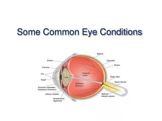
To comprehend fundamental neuro-ophthalmology, it's essential to understand measure pupil sizewith a pupilometer. This ability is necessary for eye emergencies, clinics, and, most significantly, for examinations. Pupil size determines how much light is allowed to reach the retina. It may range from around 1mm to 8mm in diameter, depending on how much light is absorbed by the retina. To feed the intrinsic muscles of the iris, the dilator and the sphincter pupillae, the sympathetic and parasympathetic neural systems interact. Patients' pupils and pupillary reflexes are critical to determining the cause of a medical condition in the clinic. ©Copyright by Neuroptics.com
Three major eye conditions that can be treated with pupil evaluation ©Copyright by Neuroptics.com
1. Adie's tonic pupil • In an Adie, the tonic pupil is an anisocoria, a condition in which the aberrant pupil is bigger and does not constrict to light but gradually constricts to accommodation. • A light-near dissociation is what is meant by this term. For lengthy periods of sustained near exertion, the pupil normally displays gradual constriction and slow re-dilatation. Damage to the parasympathetic pathway's post-ganglionic fibers is to blame. ©Copyright by Neuroptics.com
2. Acute angle-closure glaucoma • It occurs when the anterior chamber angle is mechanically constricted due to iris crowding at semi-dilated pupils. An intraocular tumor, the creation of anterior or posterior synechiae after uveitis, or rubeotic glaucoma induced by fibrovascular growth in the chamber angle due to retinal ischemia might all be possible causes (diabetes and central retinal vein occlusion classically). ©Copyright by Neuroptics.com
To confirm a diagnosis based only on history, a slit-lamp pupillary evaluationis required. The ophthalmologist should be contacted promptly if a patient has this issue. Glaucoma drops and intraocular pressure-lowering medicines may be used to decrease the eye pressure in these people, and they may be eligible for pupil measurement or an iridotomy to reduce the stress. ©Copyright by Neuroptics.com
3. Third nerve palsy • A third-nerve palsy might be fully or partially paralyzed. Third nerve palsy is characterized by a completely dilated pupil, 'down and out' eye, ptosis, and no light or accommodation constrictions. Although this is not always possible, it is possible to verify that the lesion is in the efferent route by shining light into that eye and seeing a constrictive light response in the contralateral pupil. ©Copyright by Neuroptics.com
Microvascular infarction — blockage of the vasanervorum (risks: hypertension, diabetes, atherosclerosis), a compressive lesion (aneurysm, tumor), or trauma are all possible causes of this condition. Minor third-nerve palsy symptoms may be indicative of a more severe problem. ©Copyright by Neuroptics.com