AR RM MANOS
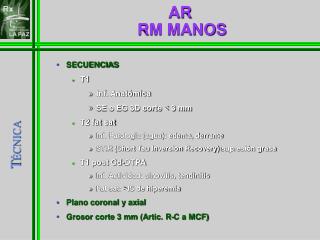
AR RM MANOS. SECUENCIAS T1 Inf. Anatómica SE o EG 3D corte < 3 mm T2 fat sat Inf. Patología (agua): edema, derrame STIR (Short Tau Inversion Recovery) supresión grasa T1 post Gd-DTPA Inf. Actividad: sinovitis, tendinitis Fat sat: >IS de hiperemia Plano coronal y axial

AR RM MANOS
E N D
Presentation Transcript
AR RM MANOS • SECUENCIAS • T1 • Inf. Anatómica • SE o EG 3D corte < 3 mm • T2 fat sat • Inf. Patología (agua): edema, derrame • STIR (Short Tau Inversion Recovery) supresión grasa • T1 post Gd-DTPA • Inf. Actividad: sinovitis, tendinitis • Fat sat: >IS de hiperemia • Plano coronal y axial • Grosor corte 3 mm (Artic. R-C a MCF) TÉCNICA
Técnica de RM (8) Lisbona MP, Maymó J. Reumatol Clin 2007; 3(3): 126-36
RM DINÁMICA • Imágenes T1 seriadas rápidas tras bolus Gd iv. • 3DEGT1 fat sat (7-10 sec de 1min a 1.2 mm) • Procesamiento de imagen, delineación manual de sinovial y sustración. Cuantificar REALCE sinovial. (9,10) • Correlación con severidad histológica de inflamación y marcadores clínicos de actividad de enf. (Tº 30-60 seg.) • Reducción vol sinovial inflamado tras Tto (6 sm) (11) • > reproducibilidad que US TÉCNICA (9) Farrant JM, et al. Skeletal Radiol 2007; 36: 269-79. (10) Reece RJ, et al. Arthritis Rheum 2002; 46: 366-72. (11) Lisbona MP, et al. Ann Rheum Dis 2005; 64:403.
EQUIPOS DE RM • ALTO CAMPO (1, 1.5 y 3T) • Mejor imagen 3T en T1 y >>T2 • Mejor: sinovitis, edema y peq. erosión CAMPO MAGNÉTICO (12) Wieners G, et al. Eur Radiol 2007; 17: 2176-82
EQUIPOS DE RM • BAJO CAMPO (<1T): E-RM (0.2, 0.3T) • Ventajas: claustrofobia, comodidad, coste (13,14,15) • Comparar AC (cte): Valorar erosiones, < realze, edema • Sinovitis: STIR (supresión grasa) <S/R < calidad • Comparar USDp en sinovitis; peor sensib, bien E (16) CAMPO MAGNÉTICO (13) Savnik A, et al. Eur Radiol 2001; 11: 1030-8. (14) Lindegaard H, et al. Ann Rheum Dis 2006; 65: 1208 (15) Schirmer C, et al. Ann Rheum Dis 2007; 66: 522-9. (16) Freeston JE, et al. Ann Rheum Dis 2008; 67: 1351.