Download
1 / 1
10 likes | 141 Views
#1448. Functional asymmetry for auditory processing in human primary auditory cortex J. T. Devlin 1 , J. Raley 1 , E. Tunbridge 1 , K. Lanary 1 , A. Floyer-Lea 1 , C. Narain 1 , P. Jezzard 1 , P. M. Matthews 1 , D. R. Moore 2

E N D
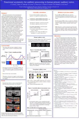
#1448 Functional asymmetry for auditory processing in human primary auditory cortex J. T. Devlin1,J. Raley1, E. Tunbridge1, K. Lanary1, A. Floyer-Lea1, C. Narain1, P. Jezzard1, P. M. Matthews1, D. R. Moore2 1Centre for Functional Magnetic Resonance Imaging of the Brain, University of Oxford, U. K. 2MRCInstitute of Hearing Research, Nottingham, U. K. Laterality calculations Relation to previous studies Summary Structural asymmetries in the supratemporal plane of the human brain are often cited as the anatomical basis for the lateralisation of language predominantly to the left hemisphere1,2. However, similar asymmetries also are found for structures mediating earlier events in the auditory processing stream3-7, suggesting that functional lateralisation may occur even at the level of primary auditory cortex. We tested this hypothesis using functional magnetic resonance imaging to evaluate human auditory cortex responses to monaurally presented tones. Relative to silence, tones presented separately to either ear produced greater activation in left than right Heschl’s gyrus, the location of primary auditory cortex. In contrast, in non-primary auditory cortex a contralateral dominance was observed. This functional lateralisation for primary auditory cortex is distinct from the contralateral dominance reported for other mammals8, including non-human primates, and may have contributed to development of the unique role for the left hemisphere in language processing. • To assess relative laterality of auditory cortex responses, we computed a Laterality Index (LI): (Contralateral - Ipsilateral BOLD signal change) (Contralateral + Ispilateral BOLD signal change) +100 indicates completely contralateral activation -100 indicates completely ipsilateral activation • Using mean signal change avoided biasing the laterality calculation through (1) an arbitrary statistical threshold for counting “active” voxels, or (2) differences in the volume of the ROIs across hemispheres (i.e. partial volume effects). • Previous MEG and fMRI studies have reported a contralateral dominance for monaural processing11-15, but have not distinguished between primary and non-primary regions. • The N1m signal in MEG is taken as an indicator of PAC but may instead reflect responses in non-primary regions lateral to PAC16,17 and thus would be consistent with the current findings. • fMRI studies have considered aggregate signal from both primary and non-primary regions. In our study, when these regions were combined, a small contralateral advantage also emerged due to the larger volume of non-primary auditory cortex. × 100 LI = Discussion Primary auditory cortex Our results demonstrate that within primary auditory cortex there is a left hemisphere dominance for processing simple auditory stimuli which is not found in surrounding, non-primary regions. If speech specific operations do not begin until the signal reaches the cerebral cortex18, then one function of PAC may be to determine whether an incoming signal has sufficient spectral and temporal complexity to be treated as speech. A left hemisphere advantage would then facilitate the rapid temporal processing in adjacent left hemisphere auditory areas19. If correct, this may explain why this pattern of functional organisation is different from the contralateral dominance seen in other species, including other primates8. On the other hand, like humans, at least two species of great apes, chimpanzees (Pan troglodyte) and gorillas (Gorilla gorilla), show structural auditory cortex asymmetries which are not seen macaques (Macaca mulatto)20-22. Thus, there appears to be a hierarchy in the degree of hemispheric lateralisation in primates, with monkeys showing the least asymmetry and humans the most. Whether the relative difference in structural asymmetry are matched by a corresponding functional shift from a contralateral auditory dominance to a left hemisphere dominance in chimpanzees and gorillas or whether left functional lateralisation for auditory processing is unique to humans remains to be demonstrated. • The majority of primary auditory cortex (PAC) is located on Heschl’s gyrus and was therefore identified on each participant’s structural scan as an anatomic correlate of PAC. E.g.: Current Study • The current study used sparse sampling9 to measure cortical auditory responses to monaurally presented tones • Participants discriminated between high (4000Hz) and low (250Hz) frequency tones (90db SPL) by pressing one of two buttons as quickly as possible after the tone onset. • Half of all trials had a silent stimulus. • The purpose of the task was simply to control attention by forcing participants to attend to the tones throughout the scanning. Figure 3: Laterality indices in PAC Left ear stimulation Right ear stimulation Figure 1: Sparse sampling paradigm 100 60 20 -20 -60 -100 100 60 20 -20 -60 -100 (p<0.001) * Expected BOLD signal Laterality index R * (p<0.01) Individual subjects Group mean (±SEM) Stimulation Paradigm Tone Tone Scanner Scanner • Within PAC, monaurally presented tones led to greater BOLD signal change in the left hemisphere regardless of which ear was stimulated 0 5 10 15 20 25 30 35 Time (seconds) Non-primary auditory cortex References 1. Geschwind, N. & Galaburda, A. M. (1985) Archives of Neurology42, 634-54. 2. Foundas, A. L., Leonard, C. M., Gilmore, R., Fennell, E. & Heilman, K. M. (1994) Neuropsychologia32, 1225-31. 3. Penhune, V. B., Zatorre, R. J., Macdonald, J. D. & Evans, A. C. (1996) Cerebral Cortex6, 661-672. 4. Rademacher, J., Caviness, V. S., Steinmetz, H. & Galaburda, A. M. (1993) Cerebral Cortex3, 313-329. 5. Geschwind, N. & Levitsky, W. (1968) Science161, 186-187. 6. Galaburda, A. M., Sanides, F. & Geschwind, N. (1978) Archives of Neurology35, 812-7. 7. Shapleske, J., Rossell, S. L., Woodruff, P. W. & David, A. S. (1999) Brain Research Reviews29, 26-49. 8. Clarey, J. C., Barone, P. & Imig, T. J. (1992) in The Mammalian Auditory Pathway: Neurophysiology (eds. Popper, A. N. & Fay, R. R.) 232-334. (Springer-Verlag, New York). 9. Hall, D. A. et al. (1999) Human Brain Mapping7, 213-223 10. Rivier, F. & Clarke, S. (1997) NeuroImage6, 288-304. 11. Jancke, L., Wustenberg, T., Schulze, K. & Heinze, H. J. (2002) Hear Res170, 166-78. 12. Pantev, C., Ross, B., Berg, P., Elbert, T. & Rockstroh, B. (1998) Audiology Neuro-Otology3, 183-190. 13. Scheffler, K., Bilicen, D., Schmid, N., Tschopp, K. & Seelig, J. (1998) Cerebral Cortex8, 156-163. 14. Suzuki, M. et al. (2002) Neuroscience Research Communications30, 197-206. 15. Woldorff, M. G. et al. (1999) Human Brain Mapping7, 49-66. 16. Liegeois-Chauvel, C., Musolino, A., Badier, J. M., Marquis, P. & Chauvel, P. (1994) Electroencephalogr Clin Neurophysiol92, 204-14. 17. Pantev, C. et al. (1995) Electroencephalogr Clin Neurophysiol94, 26-40. 18. Scott, S. K. & Johnsrude, I. S. (2003) Trends Neurosci26, 100-7. 19. Zatorre, R. J., Belin, P. & Penhune, V. B. (2002) Trends in Cognitive Science6, 37-46. 20. Gannon, P. J., Holloway, R. L., Broadfield, D. C. & Braun, A. R. (1998) Science279, 220-2. 21. Hopkins, W. D., Marino, L., Rilling, J. K. & MacGregor, L. A. (1998) Neuroreport9, 2913-8. 22. Yeni-Komshian, G. H. & Benson, D. A. (1976) Science192, 387-9. • Defined as areas adjacent to PAC which were activated by the tone vs. silence comparison in the group 1 2 3 Step 1: Tones vs. silence in RFX (cluster stats: Z>3.5, p<0.05) Step 2: Anatomically masked to include coordinates of human non-primary areas10 Step 3: Removed individual subject’s HG HG removed Masked Results • Monaural tones relative to silence activated Heschl’s gyrus and adjacent non-primary areas bilaterally Figure 2: Auditory cortex activations Figure 4: Laterality indices in non-primary areas Right hemisphere Left hemisphere PreCS Left ear stimulation Right ear stimulation IPS CS CS PreCS IPS PreCS 30 10 -10 -30 30 10 -10 -30 SMG IFS SMG PTr PT (p<0.05) (p<0.07) HG PT Insula PTr HG PP * Insula * STG PP STG STS STS MTG MTG TP Laterality index TP Left ear stimulation • In non-primary auditory cortex, monaurally presented tones led to a small contralateral advantage regardless of which ear was stimulated, and the pattern was consistent across a range of statistical thresholds for identifying group activations (Table) Right ear stimulation Table: Laterality indices in non-primary regions. Mean laterality Indices Masking threshold Left ear stimulation Right ear stimulation Z > 4.0 8 ± 3 4 ± 3 Z > 3.5 7 ± 3 9 ± 3 Z > 3.1 6 ± 3 10 ± 3 Z > 2.3 7 ±4 11 ± 5 Italics indicates non-significant effect. p<0.01 p<10-15 The upper panels display lateral views of the inflated left and right hemisphere surfaces with sulci and gyri shown in dark and light grey, respectively. The middle and bottom panels show activation in cortical auditory fields due to left and right ear stimulation.