Download

1 / 25
250 likes | 408 Views
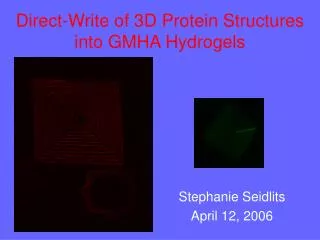
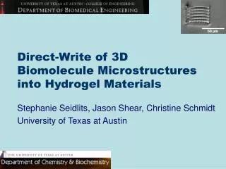
Direct-Write of 3D Biomolecule Microstructures into Hydrogel Materials. Stephanie Seidlits, Jason Shear, Christine Schmidt University of Texas at Austin. ECM Components. 3D Architecture. Alberts, MBOC, 4th ed. Bochaton-Piallat, Ophtamol Vis Sci, 2000. Engineered in vivo-like Scaffolds.

E N D
Direct-Write of 3D Biomolecule Microstructures into Hydrogel Materials Stephanie Seidlits, Jason Shear, Christine Schmidt University of Texas at Austin
ECM Components 3D Architecture Alberts, MBOC, 4th ed. Bochaton-Piallat, Ophtamol Vis Sci, 2000 Engineered in vivo-like Scaffolds Submicron Features Spatial Control Esch, J. Neurosci, 1999 Teixeira, JCS, 2003
Neuronal Cell Body Extending Neurite 3D Hydrogel Support Crosslinked Protein Structures Scaffold design
www.fsu.edu/microscopyprimer Multiphoton excitation • Advantages of MPE photocrosslinking: • Inherently 3D • Subcellular resolution • Complex geometries --possible • Heating effects minor ----and localized • Can fabricate---------------structures of natural -----materials www.cellscience.biorad.com
Resolution and biocompatibility of MPE photocrosslinking 1 µm Kaehr, PNAS, 2004
Hyaluronic acid • Enzymatically biodegradable • Photocrosslinkable --------------hydrogels (Leach, 2003, Biotech Bioeng) • Not cell adhesive • Nonimmunogenic • Angiogenesis and wound healing ring opening product hyaluronic acid glycidylmethacrylate transesterification product Adapted from Derouet, 2002, Eur Poly J
Crosslink protein structures directly into saturated GMHA hydrogels using multiphoton excitation Make GMHA hydrogels and soak in photosensitizer and protein solution Wash to remove unreacted solution MPE crosslinking in 3D
BSA-FITC Structures in Texas Red-GMHA Hydrogels Pyramid base = 50 μm
Z=40 µm Z=20 µm 50 µm 50 µm Z=0 µm 50 µm 50 µm 50 µm 50 µm 3D protein structures in HA hydrogels Z=40 µm Z=20 µm Z=0 µm
Scanning Electron Microscopy 2 µm 200 nm 2 µm
F F F F + + + + + + + B B bsa avidin Avidin-biotin functionalization Strategy 2: Strategy 1:
Avidin Structures Scale bar = 150 µm ~30 ms exposure time
50 µm 50 µm BSA structures modified with avidin ~3 ms exposure time
50 µm MPE crosslinking of proteins within 3D hydrogels creates scaffolds with: • Flexibility to incorporate multiple bioactive materials • Inherent 3D spatial control of geometry and biomolecule presentation • Submicron sized features
Acknowledgements • Christine Schmidt • Jason Shear • Rebecca Rosenberger • Kat McCarty • Jill Heisler • Curt Deister • Bryan Kaehr • Rex Nielson • Schmidt Lab Members • Shear Lab Members NSF-IGERT NSF BES-0500969 and BES-021744 Welch Foundation
IGERT: Cellular and Molecular Imaging for Diagnostics and Therapeutics The University of Texas at Austin Novel Contrast Agent Development Novel Imaging and Spectroscopy System Development Diagnostic and Vital Imaging Spectroscopy Imaging to Monitor Therapeutics Integrative Graduate Education & Research Training Program Nicholas Peppas, Sc.D., Director Jennifer Brodbelt, Ph.D., Co-Principal InvestigatorChristine Schmidt, Ph.D., Co-Principal Investigator http://www.bme.utexas.edu/igert/
A) B) Figure 4. 3D MPE crosslinked BSA structures within hydrogels. A) Optical transmission images of 200 mg/mL BSA (2% BSA-FITC) photocrosslinked in a 2 wt% GMHA hydrogel with 6 mM FAD using a pulsed TiS laser at 740 nm. Each image was taken in the same xy plane and a different z plane (the first image is focused at the top of the pyramid and the last at the bottom). C) Confocal microscope image of 200 mg/mL BSA (2% BSA-FITC) crosslinked using 6 mM FAD within a 100 mg/mL PEGDA hydrogel. Images are from different planes spanning a total depth of 80 m. Note: concentrations given are those in the uncrosslinked solution. Scale bars = 20 µm.
Crosslinking with FAD • Two Mechanisms: • Type I: triplet state abstract hydrogen to create radicals • Type II: creates singlet oxygen (Spikes 1999 Photochem Photobio) • Photocrosslinkable amino acids: Tyr, Trp, His, Cys…