Download
1 / 36
360 likes | 566 Views
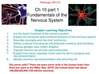
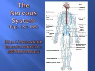
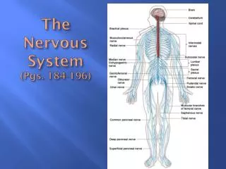
The Nervous System (Pgs. 184-196). The Nervous System. Organs of the nervous system are divided into Central Nervous System (CNS) Peripheral Nervous System (PNS). Functions of the Nervous System. Sensory Integrative Motor. Functions of the Nervous System. Sensory functions detect changes

E N D
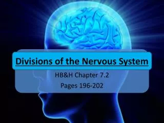
The Nervous System • Organs of the nervous system are divided into • Central Nervous System (CNS) • Peripheral Nervous System (PNS)
Functions of the Nervous System • Sensory • Integrative • Motor
Functions of the Nervous System • Sensory functions detect changes • Sensory receptors at the end of peripheral nerves detect changes inside/outside of the body • External – light, sound touch Internal – pH levels, oxygen/carbon dioxide concentrations • Information gathered is converted to nerve impulses of the PNS which sends the information to the CNS
Functions of the Nervous System • Integrative functions bring sensory information together and makes decisions that are acted upon by using motor function
Functions of the Nervous System • Motor functions are responses to sensory information • Employ peripheral nerves that carry impulses from CNS to responsive parts called effectors • Effectors are outside the NS and include a. Muscles – contract when stimulated b. Glands – secrete hormones when stimulated (glandular epithelial tissue)
Nervous Tissue • Neurons (nerve cells) are the functional unit; • Specialized to react to physical & chemical changes in their surroundings and conduct nerve impulses • Neuroglia support the physiological needs of neurons • Fig. 8-2, 8-3, 3-21 pg. 70
Anatomy of a Neuron • dendrite – provide receptive surfaces to carry impulses toward cell body; relatively short & highly branched • cell body – contains various organelles • axon – a single axon arises from cell body; transmit impulses away from the cell body; some are surrounded by specialized glia cells called Schwann cells that form myelin sheath http://www.viddler.com/explore/annette/videos/7/
Functional Differences of Neurons • motor neurons – transmit impulses form brain to an effector (efferent neurons) (multipolar) • sensory neurons – transmit impulses to spinal cord & brain (afferent neurons) (most unipolar, some bipolar) • interneurons – carry impulses from sensor neurons to motor neurons (multipolar)
Structural Differences of Neurons • bipoloar – only 2 nerve fibers, one arising from either end; found in specialized parts of the eyes, nose, & ears
Structural Differences of Neurons • unipolar/monopolar – single nerve fiber extending from its cell body; found in ganglia outside the brain or spinal cord
Structural Differences of Neurons • multipolar – many nerve fibers arising from their cell bodies; most common type of neuron in the brain & spinal cord (ganglia – a mass of neuron cell bodies, usually outside the CNS)
Neuroglia Cells • Accessory cells – Schwann cells, astrocytes, microglia, oligodendrocytes, ependymal (We will discuss the functions of each of these types of cell during the lab.) • Fill spaces, support neurons, hold nervous tissue together; play a role in the metabolism of glucose, help regulate K+ concentration, produce myelin, and carry on phagocytosis • Fig. 8-3
Regeneration of Nerve Fibers • If a neuron cell body is injured, the neuron is likely to die • If the axon of a peripheral nerve fiber is severed, its distal portion will die, but the proximal portion may regenerate & re-establish its former connections • Significant regeneration is unlikely to take place in the CNS
Structure of Peripheral Nerves – consists of bundles of nerve fibers surrounded by connective tissue – Fig. 8-4 • Epineurium – outermost layer; dense and include many collagenous fibers • Fasicicle – a bundle of nerve fibers • Perineurium – less dense connective tissue surrounding fascicle • Endoneurium – small amount of loose connective tissue that surrounds individual nerve fibers
Resting Potential • http://www.dnatube.com/video/5035/Neuron-Resting-Potential
A cell membrane is usually electrically charges or polarized so that outside is + and inside is - • Resting Potential (-70 mvolts) • Nerve cell is not conducting impulses • [Na+] is greater on the outside of the cell and [K+] is greater on the inside of the cell • There is a large number of negatively charged ions inside the cell which can’t diffuse out • At rest inside stays negative because K+ can diffuse easily out of the cell through open channels; Na+ can’t diffuse as easily into cells through “their” protein channels • Na+ /K+ pump (active transport) – maintains system so equilibrium is not reached; therefore, Na+ is always being pumped back out and K+ is being pumped back in
Local Potential Changes • Stimulation of a membrane affects its resting potential in a local region (light, temp., other neurons • Membrane starts to become depolarized (moves toward zero) • Threshold potential is reached which causes an action potential
Action Potential • http://www.dnatube.com/video/1105/Understanding-Action-Potential-and-Nerve-Impulses
Action Potential 1/1000 sec. or less • At threshold, Na+ channels open and Na+ diffuse inward causing depolarization • About the same time K+ channels open and K+ diffuses outward, causing repolarization • This rapid change in potential is an action potential • Many action potentials can occur before an active transport mechanism re-establishes the original resting potential • The propagation of actions potentials along a nerve fiber is an impulse
Refractory Period • Refractory Period • A brief time (10-30 m/sec.) following the passage of a nerve impulse when the membrane is unresponsive to ordinary stimuli • Membrane must return to resting potential before it can be stimulated again • All – or – None Response • If a nerve fiber responds at all, it responds completely • All impulses carried on that fiber will be of the same strength
Coding & Interpretation of Messages • Frequency of action potentials – a weak stimulus initiates only a few action potentials/sec., a strong stimulus initiates many (upper limit because of refractory period) • Duration of a burst of action potentials – a weak stimulus may give rise to a short burst of pulses in the neuron, a strong stimulus a longer burst • Number & kinds of neurons firing – The threshold needed to initiate a nerve impulse varies from one neuron to another. Thus a weak stimulus will cause only a few neurons to fire, strong will fire all of these neurons, plus others with higher thresholds.
Impulse Conduction • Unmyelinated fibers conduct impulses that travel over their entire surface • Myelinated – impulses travel from node to node • Impulse conduction is more rapid on myelinated fibers with large diameters • http://www.youtube.com/watch?v=DJe3_3XsBOg
The Synapse – The junction between 2 neurons. A synaptic cleft is the gap between parts of two neurons at a synapse. Fig. 8-7 • Impulses usually travel from a dendrite or cell body, then along the axon to a synapse • Axons have synaptic knobs at their distal ends that secrete neurotransmitters • The neurotransmitter is released when a nerve impulse reaches the end of an axon and the neurotransmitter diffuses across the synaptic cleft. • When the neurotransmitter reaches the nerve fiber on the distal side of the cleft, a nerve impulse is triggered.
The Synapse • http://www.dnatube.com/video/261/Neural-Synapse
Nerve Pathways – the route followed by an impulse as it travels through the nervous system, Fig. 8-5 • Reflex Arc – simplest nerve pathway • A reflex arc usually includes a sensory neuron, a reflex center composed of interneurons, and a motor neuron • Reflex arc is the behavioral unit of the nervous system • Reflex Behavior • Reflexes are automatic unconscious responses to changes • Help maintain homeostasis • Knee jerk – 2 neurons • Withdrawal reflexes are protective actions
Reflex Arc • http://www.youtube.com/watch?v=Y5nj3ZfeYDQ&feature=related