Download
1 / 21
210 likes | 624 Views
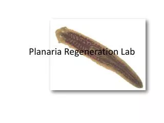
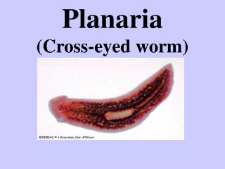
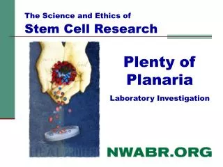
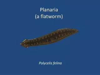
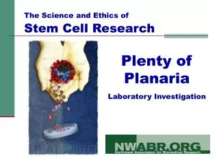
The Science and Ethics of Stem Cell Research. Plenty of Planaria Laboratory Investigation. What are Planaria?. Freshwater flatworms (phylum: Platyhelminthes) Live in freshwater under leaves and stones Avoid light (“negative phototaxis”) Free living – not parasites

E N D
The Science and Ethics of Stem Cell Research Plenty of Planaria Laboratory Investigation
What are Planaria? • Freshwater flatworms (phylum: Platyhelminthes) • Live in freshwater under leaves and stones • Avoid light (“negative phototaxis”) • Free living – not parasites • Carnivores – eat decaying meat, insects and larvae (egg yolks or liver in the lab) • 1-20 mm in size • Long lived • Use sexual or asexual reproduction This picture has been released it into the public domain.
Planaria are complex organisms Gastrovascular System
Nervous System
Reproductive System
Excretory/Osmoregulatory System
Planaria have an amazing ability to regenerate parts of themselves Planaria regeneration is a great model for demonstrating stem cell function and complexity of tissue regeneration They can grow easily in the lab Why study Planaria to learn about stem cells? 1/279th
Neoblast = Planaria Stem Cell • 20-30% of all the cells in a planaria are Neoblasts. • Neoblasts divide by mitosis and can make more of themselves. They are the only dividing cells in planaria. • When a planaria is wounded, neoblasts migrate to the site and begin dividing • Neoblasts can become ANY cell the planaria needs—nerve cells, flame cells, reproductive cells… Ann. Rev. Cell. Dev. Biol. 20:725-757, 2004
Three types of planaria with their neoblasts labeled with BrdU BrdU (bromodeoxyuridine) is substituted for thymine during DNA replication. Only dividing cells—in this case the neoblasts—will show detectible amounts of BrdU. A. Phagocata sp. B. Girardia dorotocephala C. Schmidtea mediterranea Dugesia sp. • (Dev. Biol 220: 142-153, 2000)
In Humans: Totipotent cells are limited to early embryonic division In Planaria: Neoblasts are always present Neoblasts and Stem Cells are Totipotent
Regeneration requires… turning on the right genes at the right time in the cells that come from the dividing stem cells (neoblast).
Student Laboratory Investigation Each team will receive 3 planaria: • Experimental Planaria #1 Every team will cut this into half, separating the anterior from the posterior halves. • Experimental Planaria #2 This will be cut in a manner determined by your team. • Control Planaria This will not be cut. Teacher keeps control. Used with permission from UC Museum of Paleontology
Student Laboratory Investigation • Question: What will happen when Planaria, an animal with neoblast cells, is cut in various regions of the body? • Hypothesis IF: Planaria is cut in half (transverse: horizontal), THEN: it will not die but regenerate new body parts, BECAUSE: Planaria is an animal with neoblast cells, cells that divide by mitosis and can make more of themselves (like stem cells).
Student Laboratory Investigation Materials: At the table for each pair: • 1 dissecting microscope • 1 container of pond water (share) • 1 container of Planaria (share) • 3 Petri Dishes • 2 microscope slides • 1 razor blade • 1 pipet • 1 ruler • 1 Sharpie • 1 Paint brush : Used with permission from UC Museum of Paleontology
Student Laboratory Investigation • Data: Copy chart into your lab book. Repeat for Planaria 2 and 3
Student Laboratory Investigation Procedure: • Label your petri dishes: Planaria #1,#2, Team name, period. Fill each ½ full with pond water. • Using the pipet, put one drop of pond water on slide. • Using paint brush, collect one Planaria and place on slide. Sketch, measure, and observe. • TO CUT: use 2nd slide as a platform. Use pipet or brush to transfer Planaria. • Put slide under scope and cut with blade. Sketch, measure, and observe. • Place Planaria parts into correctpetri dish. • Repeat for Planaria #2 using a different cut. • For Control (#3): follow procedures but do not cut!