Download
1 / 51
510 likes | 712 Views
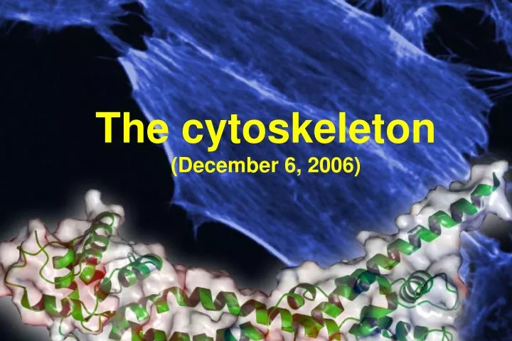
The c y toskeleton (December 6, 2006). 1. What is the cytoskeleton? 2. Filament types, and polymerization 3. Motor proteins. A dynamic framework Three types: A. Intermediate B. Microtubules C. Microfilaments. C y toskeleton.

E N D
The cytoskeleton (December 6, 2006)
1. What is the cytoskeleton? 2. Filament types, and polymerization 3. Motor proteins
A dynamic framework Three types: A. Intermediate B. Microtubules C. Microfilaments Cytoskeleton • Cellular distribution of intermediate filaments and microtubules is similar
Polimerization Three phases: 1. Lag phase: nucleation 2. Elongation 3. Equilibrium
Equilibrium 1. Dynamic equilibrium 2. Dynamic unstability: slow elongation followed by rapid (catastrophic) depolymerisation 3. ‘Tread-milling’
Torsion: Bending stiffness: Longitudinal stiffness: F F F A = persistence length F Lc = contour length Z = end-to-end distance Polymer mechanics The direction of force: Mechanism: -Intrinsic flexibility -Thermal (entropy) flexibility (persistence length)
Actin was discovered and named by a Hungarian scientist, Straub F. Brúnó
2 4 3 1 Actin monomer Globular (G-) actin MW: 43 kDa, 375 aa, 1 bound ATP or ADP Subdomains (4) nucleotide
37 nm Actin filament (F-actin) ~7 nm thick, length in vitro is more than 10 µm, in vivo 1-2 µm Double helix Semi-flexible polymer chain (persistence length: ~10 µm) "barbed end“ and "pointed end" (“barbed” =+ rapid polymerization, “pointed” =- slow polymerization)
Movement • Subcellular, cellular levels • Requires ATP (energy) • Cytoskeleton-mediated • Assembly and disassembly of cytoskeletal fibers (microfilaments and microtubules) • Motor proteins use cytoskeletal fibers (microfilaments and microtubules) as tracks
Migrating melanocyte expressing GFP-tagged actin.(Vic. SMALL).
Motility with actin polymerization Intracellular pathogens
Biophysical methods to study the cytoskeleton • Fluorescence spectroscopy • Fluorescence microscopy • Atomic force microscopy • EPR spectroscopy • Calorimetry • In vitro motility assays • …etc
Microtubuls Subunit: tubulin MW: ~50 kD, - és -tubulin -> heterodimer 1 bound GTP or GDP; a b
Microtubules ~25nm thick, tube shape 13 protofilaments Right hand, short helix Left hand, long helix Stiff polymer chain (persistence length: a few mm!) Structural polarization: + end: rapid polymerization, - end: slow polymerization GTP-cap
Tissue specific IF types Nuclear lamins A, B, C lamins (65-75kDa) Vimentin type Vimentin (54kDa) Desmin (53kDa) Peripherin (66kDa) Keratins Type I (acidic) (40-70kDa) Type II (neutral/basic) (40-70kDa) Neuronal IF neurofilament proteins (60-130kDa) The monomer is not globular, a fiber!
The subunit of filaments: „coiled-coil” dimer Vimentin dimer
protofilamentum filamentum Polymerisation of IF Polymerised in cell lack of dynamic equilibrium Central rods (-helix) hydrofob-hydrofob interactions -> colied-coil dimer 2 dimer -> tetramer (antiparallel structure) Tetramers connected longitudinally -> protofilaments 8 protofilaments -> filament
Cytoskeleton associated proteins Many families of proteins which can bind specifically to actin A. According to filaments 1. Actin-associated (e.g. myosin) 2. MT- associated (e.g. Tau protein) 3. IF- associated B. According to the binding site 1. End binding proteins („capping”, pl. gelsolin) 2. Side binding proteins (pl. tropomyosin) C. According to function 1. Cross-linkers a. Gel formation (pl. filamin, spectrin) b. Bundling (pl. alpha-aktinin, fimbrin, villin) 2. Polymerization effects a. Induce depolymerization („severing”, pl. gelsolin) b. Stabilizing (pl. profilin, tropomiozin) 3. Motor proteins
Motor proteins • They can bind to specific filament types • 2. They can travel along filaments • 3. They hydrolyze ATP
Types of motor proteins 1. Actin-based: myosins Conventional (miozin II) and nonconventional myosins Myosin families: myosin I-XVIII 2. Microtubule based motors a. Dynein Flagellar and cytoplasmic dyneins. MW~500kDa They move towards the minus end of MT b. Kinesin Cytoskeletal kinesins Neurons, cargo transport along the axons Kinesin family: conventional kinesins + isoforms. MW~110 kDa They move towards the minus end of MT 3. Nucleic acid based DNA and RNA polymerases They move along a DNA and produce force
Motor proteins • “Walk” or slide along cytoskeletal fibers • Myosin on microfilaments • Kinesin and dynein on microtubules • Use energy from ATP hydrolysis • Cytoskeletal fibers: • Serve as tracks to carry organelles or vesicles • Slide past each other
N C Common properties 1. Structure N-terminal globular head: motor domain, nucleotide binding and hydrolysis specific binding sites for the corresponding filaments C-terminal: structural and functional role (e.g. myosins) 2. Mechanical properties, function In principle: cyclic function and work Motor -> binding to a filament -> force -> dissociation -> relaxation 1 cycle requires 1 ATP hydrolysis They can either move (isotonic conditions) or produce force (isometric conditions)
ATP cycle attached ton detached toff The working cycle of motor proteins = working distance power stroke attachment detachment back stroke In vitro sliding velocity: Duty ratio: Attached time: Cycle time: d=working distance (or step size); V=ATPase activity; v=In vitro sliding velocity
Duty ratio d=working distance or step size V=ATPase activity v=in vitro motility velocity Processive motor: r->1 pl. kinesin, DNA-, RNA-polimerase the motor is attached to the track in most of the working cycle Nonprocessive motor: r->0 pl. conventional myosin A motor protein can produce force in the pN range.
How to follow polymerisation? Pyrene fluorescence filament fluorescence Monomer
The effect of Formin FH2 Dia1 Elongációs sebesség Dia3 FH2 decreased the rate of polymerisation.
F-actin mikroscop cover slit myosin In vitro motility assay
Laser tweezer Laser tweezer Micro bead Laser tweezer
Polystyrene beads of different diameters (0.5, 1, 3µm) have been functionalized with N-WASP and placed in a reconstitued motility medium containing actin, Arp2/3 complex, ADF/Cofilin, gelsolin (or any capping protein) and profilin..
A glass rod (Diam. 1µm, lenght 30 µm) has been functionalized with N-WASP and placed in the reconstitued motility medium.
Evidence for treadmilling is provided by light phase contrast recording of the movement of the rod (with A. Verkovsky): the size of the actin array remains stationnary, polymerization at rod surface being balanced by depolymerization in the actin meshwork.
A polimerizáció kémiailag befolyásolható Aktin: 1. cytochalasinok (a filamentum növekvô végéhez köt, polimerizációt gátol) 2. phalloidin (Amanita phalloides, polimert stabilizál) Mikrotubulusok: 1. Colchicin (sáfrány, őszi kikerics, antimitoticum, köszvényben ôsi idôk óta használt, MT polimerizációt gátol) 2. Vinca alkaloidok (vinblastin, vincristin, antimitoticumok, MT polimerizációt gátolnak) 3. Taxol (tiszafából, MT stabilizáló, antimitoticum)
The head group of the myosin walks toward the plus end of the actin filament it contacts.