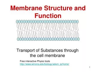
Chapter 8 Membrane Structure and Function
Chapter 8 Membrane Structure and Function. Membrane Structure. Davson-Danielli Model. Protein sandwiched on either side of a phospholipid bi-layer 8nm Surrounds cell Selectively permeable Polar heads towards the protein layer forming hydrophillic

Chapter 8 Membrane Structure and Function
E N D
Presentation Transcript
Chapter 8 Membrane Structure and Function • Membrane Structure
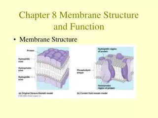
Davson-Danielli Model • Protein sandwiched on either side of a phospholipid bi-layer • 8nm • Surrounds cell • Selectively permeable • Polar heads towards the protein layer forming hydrophillic • Nonpolar tails oriented in between heads forming hydrophobic zone
Membrane Models based on research • Membranes are made of lipids. • Phospholipids can form membranes • Membrane is actually phospholipids bilayer • There is protein in membranes. • Membranes are coated on both sides with proteins
1950s electron microscopy allowed for the visibility of the plasma membrane and support the D&D Model • But………….
Problems with the D&D Model • Not all membranes are identical or symmetrical (they have different functions and are also bifacial with a distinct inside and outside face) • A membrane with an outside layer of proteins would be an unstable structure! (membrane proteins NOT soluble in water; hydrophobic regions would be in an aqueous environment)
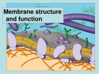
1972 Singer and Nicholson’s Fluid Mosaic Model • Fluid = wet; capable of flowing • Mosaic = an assemblage of particles • Proteins are individually embedded in bilayer • Ampipathic • Hydrophillic portions are maximally exposed to water • Hydrophobic portions in the nonaqueous environment inside the bilayer
Membranes are Fluid • Phospholipids and some proteins move laterally within the membrane • Membranes held together by weak hydrophobic attractions • Cholesterol and unsaturated hydrocarbon tails affect membrane fluidity • Saturated = no double bonds • Unsaturated = double bonds (hinder close packing of hydrocarbons)
SATURATED = less fluid/closer packing UNSATURATED = more fluid
Cholesterol modulates membrane fluidity • Less fluid at warmer temp • More fluid at cooler temp
Membrane Proteins • Integral Proteins • Embedded • Hydrophobic regions • Unilateral – reaching only partway across the membrane • Transmembrane – with hydrophobic midsections between ends exposed on both sides of membranes
Peripheral Proteins – attached to the surfaces. • Functions: transport, enzymatic activity, signal transduction, intercellular joining, cell-cell recognition, and attachment to the cytoskeleton and extra cellular matrix.
Carbohydrates • Important in cell-to-cell recognition (sorting/rejection of foreign particles) • External portions of cells • Oligosaccharides (<15 monomers) • some covalently bonded to lipids (glycolipids) • most covalently bonded to proteins (glycoproteins)