Download
1 / 53
540 likes | 606 Views
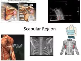
The Back and Scapular region. Dr Mukesh Singla. Muscles Connecting the Upper Limb to the Vertebral Column. Movements of Scapula. Movements of Scapula. Movements of Scapula. Movements of Scapula. Arranged in two layers Layer 1 st Trapezus Latissimus dorsi Layer 2 nd Levator scapulae

E N D
The Back and Scapular region Dr Mukesh Singla
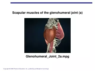
Arranged in two layers • Layer 1st • Trapezus • Latissimus dorsi • Layer 2nd • Levator scapulae • Rhomboideus major and minor
Trapezius Origin • Occipital bone(external occiptalprotuberunce), superior nuchal line, ligamentumnuchae, spine of seventh cervical vertebra, spines of all thoracic vertebrae and their supraspinous ligament Insertion • Upper fibers into posterior border of lateral third of clavicle • middle fibres- medial border of acromion and upper lip of crest of spine • lower fibers pass upward and laterally and insert on medial end of spine of scapula
Trapezius Nerve Supply • Spinal part of accessory nerve (motor) and ventral rami of C3 and 4 (sensory- proprioceptive) Action • Upper fibers along with levator scapulae elevate the scapula; • middle fibers with rhomboids pull scapula medially (retracts); • lower fibers pull medial border of scapula downward , so upper and lower fibres acting together rotate scapula-glenoid cavity face upward assisted by lower 5 digitations of serratus anterior
Latissimus dorsi Origin • Iliac crest, lumbar fascia, spines of lower six thoracic vertebrae(T7-T12), lower three or four ribs, and inferior angle of scapula (5) Insertion • Floor of bicipital groove of humerus (1) Nerve Supply • Thoracodorsal nerve • C6, 7, 8, Action • Extends, adducts, and medially rotates the arm (3) • Its called the climbing muscle • Raising of the trunk above the arm
Levator scapulae Origin • Transverse processes of first fourth cervical vertebrae (1) Insertion • Medial border of scapula (1) Nerve supply • C3 and 4 and dorsal scapular nerve • C3, 4, 5 Action • Raises medial border of scapula
ligamentum nuchae The ligamentum nuchae is a large median ligament composed of tendons and fascia located between the posterior muscles of the neck. It covers the spines of C1 to C6 vertebrae. It is a superior and posterior extension of the supraspinous ligamen
Rhomboid minor Origin • Ligamentumnuchae and spines of seventh cervical and first thoracic vertebrae (3) Insertion • Medial border of scapula (1) Nerve supply • Dorsal scapular nerve C4, 5
Rhomboid major Origin • Second to fifth thoracic spines Insertion • Medial border of scapula (1) Nerve supply • Dorsal scapular nerve C4, 5 Action • Retract scapula • The rhomboids work collectively with the levator scapulae muscles to elevate the medial border of the scapula, downwardly rotating the scapula with respect to the glenohumeral joint.
Deltoid Origin • Lateral third of clavicle, acromion, spine of scapula Insertion • Middle of lateral surface of shaft of humerus Nerve supply • Axillary nerve C5, 6 Action • Middle fibers Abducts arm; anterior fibers flex and medially rotate arm; posterior fibers extend and laterally rotate arm • Abduction from 15-90 degrees
Structures under cover of Deltoid BONE- Upper part humerus, coracoid process ,greater tubercle, lesser tubercle Intertuberculus sulcus Bursa- Subdeltoid , subacromial bursa Muscles – attached around shoulder joint Vessel- Ant and Post circumflex humeral vessel Nerves- Axillary
Supraspinatus Origin • Supraspinous fossa of scapula Insertion • Greater tuberosity of humerus; capsule of shoulder joint Nerve supply • Suprascapular nerve 5, 6 Action • Abducts arm and stabilizes shoulder joint • Initiation of abduction 0-15 degrees
Infraspinatus Origin • Infraspinous fossa of scapula Insertion • Middle impression of greater tubercle of humerus; capsule of shoulder joint Nerve supply • Suprascapular nerve after passing through spino-glenoid notch 5, 6 Action • Laterally rotates arm and stabilizes shoulder joint
Teres major Origin • Lower third of lateral border of scapula Insertion • Medial lip of bicipital groove of humerus Nerve supply • Lower subscapular nerve C6, 7 Action • Medially rotates and adducts arm and stabilizes shoulder joint
Teres minor Origin • Upper two thirds of lateral border of scapula Insertion • Greater tuberosity of humerus; capsule of shoulder joint Nerve supply • Axillary nerve (C4), C5, 6 Action • Laterally rotates arm and stabilizes shoulder joint
Subscapularis Origin • Subscapularfossa Insertion • Lesser tuberosity of humerus Nerve supply • Upper and lower subscapular nerves C5, 6, 7 Action • Medially rotates arm and stabilizes shoulder joint
Rotator Cuff • The rotator cuff is the name given to the tendons of the subscapularis, supraspinatus, infraspinatus, and teres minor muscles • are fused to the underlying capsule of the shoulder joint • stabilizing the shoulder joint • The cuff is deficient inferiorly, and this is a site of potential weakness.
Axillary Nerve • The axillary nerve arises from the posterior cord of the brachial plexus (C5 and 6) in the axilla • It passes backward and enters the quadrangular space with the posterior circumflex humeral artery • As the nerve passes through the space, it comes into close relationship with the inferior aspect of the capsule of the shoulder joint and with the medial side of the surgical neck of the humerus • It terminates by dividing into anterior and posterior branches
Axillary Nerve • branches: • An articular branch to the shoulder joint • An anterior terminal branch, which winds around the surgical neck of the humerus beneath the deltoid muscle; it supplies the deltoid and the skin that covers its lower part.
Axillary Nerve • A posterior terminal branch, which gives off a branch to the teres minor muscle and a few branches to the deltoid, then emerges from the posterior border of the deltoid as the upper lateral cutaneous nerve of the arm • The axillary nerve can be injured in dislocations of the shoulder joint
Arterial Anastomosis Around the Shoulder Joint • The extreme mobility of the shoulder joint may result in kinking of the axillary artery and a temporary occlusion of its lumen • To compensate for this, an important arterial anastomosis exists between the branches of the subclavian artery and the axillary artery • ensuring that an adequate blood flow takes place into the upper limb irrespective of the position of the arm
Arterial Anastomosis Around the Shoulder Joint Branches from the Subclavian Artery • The suprascapular artery, which is distributed to the supraspinous and infraspinous fossae of the scapula • The superficial or transverse cervical artery, which gives off a deep branch that runs down the medial border of the scapula Branches from the Axillary Artery • The subscapular artery and its circumflex scapular branch supply the subscapular and infraspinousfossae of the scapula, respectively. • The anterior circumflex humeral artery • The posterior circumflex humeral artery • Both the circumflex arteries form an anastomosing circle around the surgical neck of the humerus
Triangle Of Auscultation Boundaries- Below- Horizontal fibres of latissimus dorsi Medially- lateral border of trapezius Laterally- vertebral border of scapula Floor – 6th and 7th rib Apex of lower lobe of both lungs lie beneath this triangle
MCQ • Injury to which of the following nerve leads to winging of scapula • Long thoracic nerve • Thoracodorsal nerve • Suprascapular nerve • Dorsal scapular nerve