Download

1 / 48
480 likes | 523 Views
Learn about uterine cancer, including risk factors, types, symptoms, diagnosis methods, and histological subtypes. Explore clinical stages, differential diagnosis, and metastasis routes to enhance awareness and knowledge.

E N D
Uterine Cancer Xi-Shi Liu Obstetrics and Gynecology Hospital Fudan university 2012.08
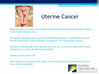
General Description • Uterine cancer is one of the most common malignancy of female genital tract. • The incidence is increasing worldwide in recent years. • Overall,2%-3% of women develop uterine cancer during their lifetime.
General Description • A malignant epithelial disease that occurs in endometrial gland of uterus • Also called endometrial cancer
Classification(pathogenetic,biologic behavior ) • Estrogen dependent type • have a history of exposure to unopposed estrogen (either endogenous or exogenous). • Hyperplastic endometrium • Better differentiafed • ER(+),PR(+) • Mere favorable prognesis
Estrogen independent type -- Have no source of estrogen stimulation of endometrium. --Arising in background of atrophic endemetrium --Less differentiated --ER(-)PR(-) --Poor prognosis
Risk Factors 1. Medical conditions a. Diabetes mellitus, hypertension. b. Overweight---obesity (excess estrogen as a result of peripheral conversion of adrenally derived androstenedione by aromatization in fat). c. Late menopause.
Risk Factors 2. Some gynecologic diseases ( Long-term endogenous estrogen exposure ) - polycystic ovary syndrome - functioning ovarian tumors - anovulating dysfunctional bleeding - Infertility, Nulliparity.
Risk Factors 3. Prolonged Use of estrogen a. Prolonged menopausal estrogen replacement therapy without progestogen. b. Prolonged use of the antiestrogen tamoxifen for breast cancer.
Risk Factors 4. Genetic factors and other factors a. Endometrial and ovarian cancer are the simultaneously occurring with other genital malignancy ,reported incidence (1.4~3.8%). b. Family history of tumor is higher.(12-28%)
Five histological subtypes • Endometrioid adenocarcinoma • Mucinous carcinoma • Serous adenocarcinoma • Clear cell carcinoma • Other rare subtypes
Five histological subtypes--Endometrioid Adenocarcinoma • Account for about 80~90%. • Well differentiated. • Prognosis is better.
Five histological subtypes--Mucinous carcinoma Rare (about 5%) a. Most of them is a well differentiated. b. Behavior is similar to that of common endometrial carcinoma.
Five histological subtypes--Serous adenocarcinoma a. Architecture is identical with complex papillary. b. More aggressively with deep myometrial and lymphatic invasion. c. Simulating the behavior of ovarian carcinoma.
Five histological subtypes--Clear cell carcinoma a. A rare subtype b. Is high grade and aggressive c. Prognosis is similar to or worse than that of papillary serous carcinoma d. Survival rate is lower 33%~64%
Five histological subtypes--other rare subtypes • Squamous adenocarcinoma • Undifferentiated carcinoma • Mixed adenocarcinoma
Clinical Features--Symptoms • Asymptomaic (about less than 5% ) • Abnormal vaginal bleeding (premenopausal or postmenopausal, minimal or nonpersistant) • Abnormal vaginal discharge(25% infection of uterine contents) • Pelvic pressure or discomfort (uterine enlargement or extrauterine disease spread)
Clinical Features--Signs • No evidence in early stage on physical examination • Slight enlargement of uterine size and soft • Uterus fixed, immobile, adenexal mess in advanced stage
Special Examination Dilation and fractional curettage ( D. C) • Most effective ,definitive procedure and commonly used • Significance -Established correct diagnosis, clinical stage -differentiated from cervical cancer or cervical involvement
Ultrasonography • Useful adjuvant method • Significances • Size of lesion • Invasion of endometrium or cervix • Resistant index of new vessels
Endometrial carcinoma in a 58-year-old woman with substantial postmenopausal bleeding. (A) Sagittal transvaginal US scan shows the endometrium with a thickness of 44 mm and a large area of mixed echogenicity suggestive of a mass. (B) Transverse sonohysterogram shows a 50-mm-diameter polypoid mass protruding into the endometrial cavity (calipers indicate the stalk of the mass). Histopathologic findings indicated poorly differentiated endometrial carcinoma. B A
Hysteroscopy • Significance -Direct observation -Taking sample correctly -Identifying polyps and submucous myoma
Pap test • -Unreliable diagnostic test • -30%-50% abnormal pap test results • Others • -MRI, CT, chest x-ray, IV urography, cystoscopy, sigmoidoscopy,
Diagnosis • History, and clinical sign , related risk factors symptoms • Diagnostic methods
Differential Diagnosis • Senile endometritis / vaginitis • Dysfunctional uterine bleeding • Submucous myoma / Endometrial polyps • Cervix cancer / Sarcoma of uterus/ Primary carcinoma of fallopian tube
Metastasis Route • Direct extension • Lymphatic metastasis: important route • Hematogenous metastasis
Clinical Stage(FIGO 1971) • Stage I Ia The carcinoma is confined to the corpus and the length of the uterine cavity is ≤ 8 cm Ib The carcinoma is confined to the corpus and the length of the uterine cavity is > 8 cm • Stage IIThe carcinoma has involved the corpus and the cervix, but has not extended outside the uterus
Clinical Stage(FIGO 1971) • Stage IIIThe carcinoma has extended outside the uterus, but not outside the true pelvis • Stage IV IVa The carcinoma has extended outside the uterus and involves the mucosa of the bladder or rectum (a bullous oedema as such does not permit the case to be allotted to Stage IV) IVb The carcinoma has extended outside the true pelvis and spread to distant organs
Surgical pathologic staging (FIGO 1988) • Stage I Ia* Tumour limited to the endometrium Ib* Invasion to less than half of the myometrium Ic* Invasion equal to or more than half of the myometrium • Stage II IIa* Endocervical glandular involvement only IIb* Cervical stromal invasion
Surgical pathologic staging (FIGO 2000) • Stage III IIIa* Tumour invades the serosa of the corpus uteri and/or adnexae and/or positive cytological findings IIIb* Vaginal metastases IIIc* Metastases to pelvic and/or para-aortic lymph nodes • Stage IV IVa* Tumour invasion of bladder and/or bowel mucosa IVb* Distant metastases, including intra-abdominal metastasis and/or inguinal lymph nodes
Stage Ia* Tumor limited to the endometriumStage Ib* Invasion to less than half of the myometriumStage Ic* Invasion equal to or more than half of the myometrium
Stage IIa* Endocervical glandular involvement onlyStage IIb* Cervical stromal invasion
Stage IIIa* Tumor invades the serosa of the corpus uteri and/or adnexae and/or positive cytological findingsStage IIIb*Vaginal metastasesStage IIIc* Metastases to pelvic and/or para-aortic lymph nodes
Stage IVa* Tumor invasion of bladder and/or bowel mucosaStage IVb* Distant metastases, including intra-abdominal metastasis and/or inguinal lymph nodes
Treatment • Surgery Radiation • Chemotherapy Hormone therapy • Early stage --- surge+ postoperative adjuvant therapy • Advanced stage --- radiation+ surge+ medicine
Principle of choice • General condition (Age, complication) • Clinical stage • Tumour pathologic type
Surgery • Object • Operative pathologic stage, finding prognosis risk factors • Remove uterus and metastasis tumour • Stage I : • Abdorminal hysterectomy + bilateral salpingoophorectomy + selective lymphadenectomy • clear cell or papillary carcinoma– omentectomy+appenditectomy
Stage II • Radical hysterectomy + pelvic lymphadenectomy + paraortic lymphadenectomy • Stage III,IV • Cytoreductive surgery
Indications of pelvic lymphadenectomy • Special pathogenetic pattern • Endometrial cancer, grade 3 or no differentiation • Myo-invasion more than ½ • Size of lesion more than 50% of uterine cavity • Involvement in isthmus of uterus
Radiation therapy • Radiation alone • Radiation with surgery
Radiation combined surgery--Radiation after surgery • Adenexal / serosal / parametrial spread • Vaginal metastasis • Lymph node metastasis • Intraperitoneal spread • Bladder / rectal invasion • Myoinvasion > 50% • G3 < 50% myoinvasion
Indications for radiation alone • Elderly or obesity • Multiple chronic or acute medical illness (hypertension, cardial disease, diabetes, pulmonary, renal) • Advanced stage unsuitable for surgery
Hormone Therapy • mechenism • Most endometrial cancers have both ER & PR.(Estrogen dependent subtype) • Indications: • Advanced or recurrent stage • Early stage and desire for fertility • Used drugs • MPA
Chemotherapy • Advanced stage or recurrent carcinoma • Postoperative adjunctive treatment for high risk factor • Used drugs: • DDP (cisplatin), CTX (cyclophosphamide), ADM (doxorubicin ), 5-Fu,Taxal MMC, VP16.
Prognostic Factors • Tumour bilologic bihavior • Cell type • Histological grade • Depth of myometrium infiltration • lymph-node metastasis • Presence of lymph vascular space involvement • Positive peritoneal cytology • General condition • Old age • Acute or chronic medical illness • Choice of treatment
5-Year Survival Rate • Stage I b: 94% • Stage I c: 87% • Stage II : 84% • Stage III : 40-60%
Follow-up • 75-95% disease will recur within 2-3 years after operation. • Items • Main complaints • Pelvic examination • Vaginal discharge smear • Chest X ray • Serum CA125 • Blood routine test • Blood biochemistry examination • CT/MRI
Questions • How to make diagnosis of uterine cancer? • What’s the principle of treatment on patients with uterine cancer? • What’re associated with prognosis of uterine cancer? ?
THANKS THE END