Download
1 / 26
320 likes | 575 Views
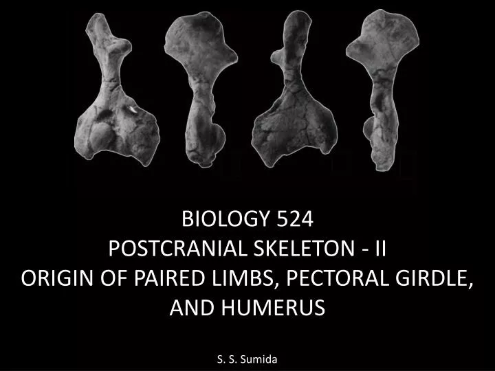
BIOLOGY 524 POSTCRANIAL SKELETON - II ORIGIN OF PAIRED LIMBS, PECTORAL GIRDLE, AND HUMERUS S. S. Sumida. Major themes we examine in this lecture include : The origin of paired limbs in general. Evolution of the pectoral girdle: detachment of girdle from skull

E N D
BIOLOGY 524 POSTCRANIAL SKELETON - II ORIGIN OF PAIRED LIMBS, PECTORAL GIRDLE, AND HUMERUS S. S. Sumida
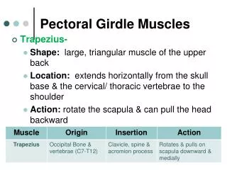
Major themes we examine in this lecture include: • The origin of paired limbs in general. • Evolution of the pectoral girdle: detachment of girdle from skull • Evolution of the pectoral girdle: reduction in emphasis on dermal elements. • Evolution of the pectoral girdle: increase in emphasis on endochondral elements • Humerus: evolution of orientation and major processes. • Change in orientation and posture of gleno-humeral joint
Embryology: All elements of the pectoral girdle, as well as the humerus are all derived from mesoderm. The pectoral girdle is a composite structure made of multiple elements, some of which form intramembranously, and some of which form endochondrally. The humerus forms endochondrally as well.
FIN FOLD THEORY OF THE ORIGIN OF PAIRED FINS In the FIN FOLD THEORY, it is hypothesized that primitive fishes (probably gnathostomes) may have had a continuous , laterally directed stabilizing structure one each side of the body. It is further hypothesized that the pectoral (fore) and pelvic (hind) fins were pieced out of these elongate, trans-segmental structures.
This would then suggest that the earliest fins were BROAD-BASED FINS with multiple basals. This theory is supported by the fact that there are primitive fishes known as Arthrodires that have a series of spines in exactly that lateral position, and some feel they may have helped to support a lateral fin fold. For example, the arthrodireClimatus:
Additionally, some extinct Paleozoic sharks (Cladoselache and its relatives) have very broadly based fins, so we know that the proposed anatomical model is a viable one. Eventually, the articulations of both the pectoral and pelvic limbs become more restricted, with a single element (humerus or femur) articulating with either girdle . This is known as a MONOBASAL ARTICULATION.
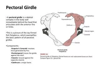
FISHES: PECTORAL GIRDLE AND FIN Four main features must be noted about the pectoral girdles of most fishes: They are physically attached, articulated with the back of the skull; and The are composed primarily of intramembranous (dermal) elements. These elements include postopercular bones, the SUPRACLEITHRUM, CLEITHRUM, and CLAVICLE. The only endochondral element is the SCAPULOCORACOID. However, it is worth noting that the scapulocoracoid provides the articulation for the humerus.
As basal tetrapods diverged from crossopterygian fishes, two significant changes took place: The pectoral girdle detached from the back end of the skull. Although dermal elements remain prominent, endochondral elements become more and more important and greater in volume.
Ventastega is considered one of the most primitive known of all tetrapods. Note the larger scapulcoracoid, and the presence of a new dermal element: the INTERCLAVICLE.
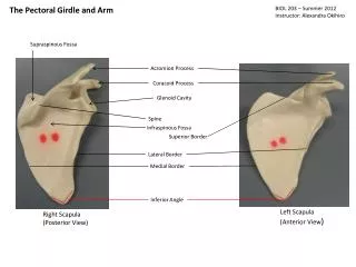
TRENDS IN THE TETRAPOD PECTORAL GIRDLE • Through the course of tetrapod evolution through to basal amniotes: • the anocleithrum is lost • the supracleithrum is reduced drastically or lost • the cleithrum is reduced significantly • the clavicle remains as the most robust dermal element left • the interclavicle remains as well. • The scapula and coracoid are recognizable as distinct elements • Perhaps the most significant anatomical distinction is the that glenoid fossa becomes very complicated in its shape, as it matches the complexly shaped humeral head and provides a track in which that element is guided.
ADDITIONAL REPTILIAN PECTORAL DIVERSITY In other reptilian groups, notice the variation in the scapula and coracoid (Ac). Note particularly the extreme reduction in elements (and increase in axial sternum) in birds.
THE PECTORAL GIRDLE: THE ROAD TO MAMMALS • The change from a basal amniote condition as seen in pelycosaurs to that of mammals is marked by the following: • Continued reduction and eventual loss of the cleithrum. • Reduction of the clavicle, and loss in cursorial mammals. • Gradual reduction in the contribution of the coracoid until is a process fused to the scapula.
HUMERUS A distinct humerus become apparent in the crossopterygian sister-taxa to tetrapods. In advanced crossopterygians, the first element of the monobasal articulation can be identified as a distinct humerus.
As the humerus becomes distinct in Devonian crossopterygian and primitive tetrapods, it becomes approximately “L-shaped”. • This very large process is the ENTEPICONDYLE (labeled “4” above), and is for attachment of progressively stronger flexor muscles. • It has been suggested that when the arm reaches forward, very strong flexors of the elbow wold be required to pull the animal forward past the planted manus.
THE TETRAPOD HUMERUS The humerus in basal tetrapods is extremely complex in construction, initially bearing little resemblance to that of more familiar recent mammals and birds. It has been various described at a “tetrahedron”, a pair of tetrahedrons mounted at an angle ton one another, and others. Following are illustrations and photographs of the diadectomorphDiadectes, and the basal reptile Labidosaurus respectively.
There are numerous things to notice about the amphiban / amniotehumerus: • Proximally the deltoid, supinator, and pectoral processes are very powerfully developed. • There is little or no distinct shaft in more basal taxa. • As in basal tetrapods, the entepicondyle is very highly developed, highlighting the continued importance of the flexor musculature of the forearm. • Distally, the ECTEPICONDYLE become well developed as well, for attachment of the extensor musculature of the forearm. • Also distally, the first structures on the road to mammals are a clearly developed spherical capitulum for reception of the radius, and trochlear notch for reception of the ulna.
THE TETRAPOD GLENOHUMERAL JOINT • With the unusual shapes of both the glenoid fossa and the proximal articualar head of the humerus in mind, their interaction must be addressed. • The basal tetrapod glenoid is not a simple concave socket. • It is generally characterized as a “SCREW-SHAPED STRAP”. • On the next page, note that the anterior part of the glenoid has a shelf above so that the articular surface faces ventrally; whereas toward the posterior part of the glenoid the articular surface faces up. • THIS FORCES THE HEAD OF THE HUMERAL TO TRACK THROUGH THIS SET OF SURFACE, ROTATING FROM FACING DOWN TO UP, EFFECTIVELY CAUSING THE HUMERUS TO ROTATE ON ITS LONG AIXS AS IT RETRACTS.
THE TETRAPOD GLENOHUMERAL JOINT • With the unusual shapes of both the glenoid fossa and the proximal articualar head of the humerus in mind, their interaction must be addressed. • The basal tetrapod glenoid is not a simple concave socket. • It is generally characterized as a “SCREW-SHAPED STRAP”. • On the next page, note that the anterior part of the glenoid has a shelf above so that the articular surface faces ventrally; whereas toward the posterior part of the glenoid the articular surface faces up. • THIS FORCES THE HEAD OF THE HUMERAL TO TRACK THROUGH THIS SET OF SURFACE, ROTATING FROM FACING DOWN TO UP, EFFECTIVELY CAUSING THE HUMERUS TO ROTATE ON ITS LONG AIXS AS IT RETRACTS.
MORE DERIVED HUMERI • In mammals: • A distinct shaft becomes much more apparent. • The proximal articular surface becomes the partially spherical humaral head, adopting a much simpler shape and greater range of motion. • The deltoid and pectoral processes unite to become the delto-pectoral crest. • The degree of development of various processes is dependent on the lifestyle of the particular mammal. • Humeral morphology in mammals is much more variable than in birds, as that of birds is significantly constrained by the requirements for flight.