Download
1 / 70
700 likes | 781 Views
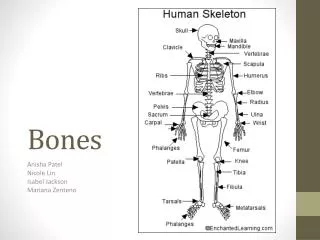
BONES. 206 Bones are said to be in the Human Skeleton. Although the human skeleton is initially made up of cartilages and fibrous membranes this is soon replaced by bone. Bones Basic Structure, Types, and Locations.

E N D
BONES • 206 Bones are said to be in the Human Skeleton. • Although the human skeleton is initially made up of cartilages and fibrous membranes this is soon replaced by bone.
BonesBasic Structure, Types, and Locations • When discussing the skeleton is it essential to review over the types of cartilages found in the body. • Hyaline Cartilages • Elastic Cartilages • Fibrocartilages
Cartilage • Hyaline Cartilage- Provides support with flexibility and resilience. Makes up the following in the human skeleton • 1. articular cartilage- covers the ends of most bones • 2. costal cartilage- connect ribs to sternum • 3. respiratory cartilage – found in make up larynx • 4. nasal cartilage- which support the external nose.
Cartilage • Elastic Cartilage- Very much like hyaline cartilage but ALSO contains more stretch elastic fibers and better able to stand repeated bending. • Makes up only 2 skeletal locations • 1. external ear- • 2. epiglottis- flap that covers the opening to the larynx each time we swallow.
Cartilage • Fibrocartilages Cartilage- are highly compressible and have great tensile strength. It is in between hyaline and elastic cartilages • Found in sites that are subjected to both heavy pressure and stretch. • 1. Menisci- padlike cartilages of the knee • 2. Disk between the vertebrae.
Always remember bone and cartilage are always distinct different tissues. Bone has a hard matrix while cartilage has a flexible matrix that can accommodate mitosis. Cartilage
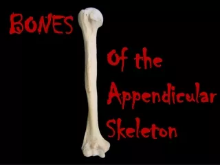
Axial Skeleton- consists of the bony and cartilaginous parts that support and protect the organs of the head, neck and trunk. Appendicular Skeleton- consists of the bones of the upper and lower limbs and bones that anchor the limbs to the axial skeleton. Division of the Skeleton
Bone Structure • Bones differ in size and shape but have similar structure, development, and functions.
Bone Classification Long Bones • Long longitudinal axes, and expanded end. • Ex: Forearm and thigh bones.
Bone Classification Short Bones • Cube like, with lengths and widths roughly equal . • Bones in the wrists and ankles.
Bone Classification Flat Bones • Plate like structure with broad surfaces • Ex: Ribs, scapulae, and some bones of the skull.
Bone Classification Irregular Bones • Variety of shapes and are usually connected to several other bones. • Ex: Vertebra, and some facial bones.
Bone Function • Bones shape, support, and protect body structures. • They also act as levers. • House tissue that produces bone/blood cells • Stores various inorganic salts.
Bone Function • Support- all the softer tissues of the body; they literally hang from the skeletal framework. • Protection- hard, bony “boxes” protect the delicate structures within them. Example: skull to brain • Movement- Muscles are anchored firmly to bones.
Bone Function • Mineral and Growth Factor Storage- Bone is a reservoir for minerals like calcium and phosphate. It also stores important growth factors. • Blood Cell Formation- Most blood formation or hematopoiesis occurs in the marrow cavities of certain bones.
Bone Structure • Because bones contain tissue they are considered an organ • They contain not only bone (osseous) tissue but nervous tissue, connective tissue, muscle tissue, and epithelial tissue.
Bone Marking • Throughout the next month as you work with this bones you will need to recognize these markings during lab • Bones are rarely smooth..they display projections, depressions, and openings that serve as sites of muscle, ligament, and tendon attachment as joint surfaces or conduits for blood vessels and nerves.
Projections (bulges) that grow outward from the bone surface includes Heads, trochanters, spines, and others. These are just a few a complete list is on pg 179 Bone Marking
Bone depressions and opening include Fossae, sinuses, foramina, and grooves These are just a few a complete list is on pg 179 Bone Marking
Bone Texture • When discussing bone texture the outward appearance has a smooth solid look to the naked eye and is referred to as Compact Bone • The internal layer is referred to as Spongy Bone and looks like a honeycomb. • We will discuss this in more detail later.
Structure of a Long Bone • Diaphysis- Shaft of the bone, located between epiphysis. A hollow tube made of hard compact bone, hence a rigid and strong structure light enough in weight to permit easy movement. • It surrounds a central medullary cavity…”Marrow cavity” • In adults the medullary cavity contains fat (yellow marrow) and is called yellow bone marrow cavity.
Structure of a Long Bone • Epiphysis- • The outer ends (joints) of a long bone. • The exterior of epiphysis is compact bone while the interior of epiphysis is spongy bone. • Outer portion of the epiphysis is coated with a layer of hyaline cartilage called Articular Cartilage. Functions like a small rubber cushion . • Red bone marrow fills in small spaces in the spongy bone composing the epiphyses.
Structure of a Long Bone • Epiphysis • Between the epiphysis and diaphysis of an adult long bone is an epiphyseal line and remnant of the epiphyseal plate. • The epiphyseal plate is a disc of hyaline cartilage that grows during childhood to lengthen the bone.
Structure of a Long Bone • Another structure in all long bones are membranes • Peristeum- a strong fibrous membrane covering a long bone except at joint surfaces, where it is covered by articular cartilage. • A thin membrane containing bone-forming cells called Endosteum lines the internal bone surfaces.
2 Types of Bone • Compact Bone- Wall of the diaphysis is mainly composed of this. Hard and dense; continues matrix with no gaps. • Spongy Bone- Found on the ends of the epiphysis…consists of many branching bony plates. Contains spaces that may be filled with marrow. The needle-like threads of spongy bone that surround a network of spaces are called trabeculae.
In Compact Bone the matrix is organized into numerous structural units called osteons or Haversian systems. Each circular and tube like osteon is composed of calcified matrix. The rings are called a concentric lamella. Microscopic- Anatomy of Bone
Draw and label the long bone on pg. 180 in book. http://www.mhhe.com/biosci/ap/holeessentials/student/olc/matching0160.html http://kidshealth.org/kid/body/bones_SW.html Parts of the Long Bond
Bone Development • Ossification and Osteogenesis are synonyms meaning the process of bone formation. • In embryos this process leads to the formation of the bony skeleton • And continues on until early adulthood as the body continues to grow in size.
Bone Development • The first 8 weeks of development the human embryo is completely fibrous membranes and cartilage • Bones continue to grow and develop into adulthood. • Bones form by replacing existing connective tissue in one of two ways • Intramembranous Bones and Endochondral Bones
INTRAMEMBRANOUS BONES • When a bone develops from a fibrous membrane it is called intramembranous ossification and the bone is called a membrane bone. • Intramembranous ossification results in the formation of Flat Bones (Mostly of the skull and clavical). • During development membrane like layers of connective tissues appear at the area of future bones. • Layers supplied with blood vessels and tissue arranged around the vessels • Cells enlarge and change into bone-forming tissue called OSTEOBLAST.
ENDOCHONDRAL BONES • Ex: Most all bones • They develop as hyaline cartilage that is later replaced by bone tissue. • This is more complex than intramembranous ossification because the hyaline cartilage must be broken down as ossification proceeds • Primary ossification centers appears in the diaphysis, whereas secondary ossification centers appear in the epiphyses • An epiphyseal plate remains between the primary and secondary ossification centers. Development proceeds from masses
Osteo….The different types bone cells. • Osteoblasts - make new bone and help repair damage; • Osteocytes- mature bone cells.carry nutrients and waste products to and from blood vessels in the bone; • Osteoclasts- break down,reabsorb bone and help to sculpt and shape it. Osteoclasts are very active in kids and teens, working on bone as it is remodeled during growth. They also play an important role in the repair of fractures.
An epiphyseal plate consists of layers of cells: resting cells, young cells, older enlarging cells and dying cells. The epiphyseal plates are responsible for lengthening. Long bones continue to lengthen until the epiphyseal plates are ossified. Bone Growth- Epiphyseal Plate
Growth in thickness is due to intramembranous ossification beneath the periosteum. The action of osteoclasts forms the medullary cavity. Bone Growth- Epiphyseal Plate
HOMEOSTASIS OF BONE TISSUE • Osteoclast and osteoblast continually remodel bone. • The total mass of bone remains nearly constant throughout life. While it may appear to be lifeless it is VERY active • Adult skeleton normally recycles 5-7% of our bone mass a week…spongy bone replaced every 3-4 years, compact bone every 10.
Sunlight, hormonal secretions, and exercise all affect bone development Deficiencies of vitamin A,C, or D result in abnormal development. Physical stress Exercise thickens and strengthens bone tissue Lack of activity can cause waste and thin tissue. Factor Affecting Bone Development
BONE- Blood Cell Formation • Hemopoiesis- the process of blood formation. • Begins in the yolk, which lies outside the embryo. Later in development, blood cells are manufactured in the liver, spleen, and still later they form in the bone marrow.
BONE- Blood Cell Formation • Marrow- soft tissue within the medullary cavity of long bones, in spongy bone, and in canals of compact bone tissue.