Figure 13.6
Cervical plexus. Cervical nerves C 1 – C 8. Brachial plexus. Cervical enlargement. Thoracic nerves T 1 – T 12. Intercostal nerves. Lumbar enlargement. Lumbar nerves L 1 – L 5. Lumbar plexus. Sacral plexus. Sacral nerves S 1 – S 5. Cauda equina. Coccygeal nerve Co 1.
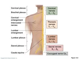

Figure 13.6
E N D
Presentation Transcript
Cervical plexus Cervical nerves C1 – C8 Brachial plexus Cervical enlargement Thoracic nerves T1 – T12 Intercostal nerves Lumbar enlargement Lumbar nerves L1 – L5 Lumbar plexus Sacral plexus Sacral nerves S1 – S5 Cauda equina Coccygeal nerve Co1 Figure 13.6
Gray matter White matter Dorsal and ventral rootlets of spinal nerve Ventral root Dorsal root Dorsal root ganglion Dorsal ramus of spinal nerve Ventral ramus of spinal nerve Spinal nerve Rami communicantes Sympathetic trunk ganglion Anterior view showing spinal cord, associated nerves, and vertebrae. The dorsal and ventral roots arise medially as rootlets and join laterally to form the spinal nerve. Figure 13.7 (a)
Dorsal ramus Ventral ramus Spinal nerve Rami communicantes Intercostal nerve Dorsal root ganglion Sympathetic trunk ganglion Dorsal root Ventral root Branches of intercostal nerve • Lateral cutaneous • Anterior cutaneous Sternum (b) Cross section of thorax showing the main roots and branches of a spinal nerve. Figure 13.7 (b)
Cervical Plexus • Formed by ventral rami of C1–C4 • Innervates skin and muscles of the neck, ear, back of head, and shoulders • Phrenic nerve • Major motor and sensory nerve of the diaphragm (receives fibers from C3–C5)
Ventral rami Segmental branches Hypoglossal nerve (XII) Ventral rami: Lesser occipital nerve C1 Greater auricular nerve C2 Transverse cervical nerve C3 Ansa cervicalis C4 Accessory nerve (XI) C5 Phrenic nerve Supraclavicular nerves Figure 13.8
Brachial Plexus • Formed by ventral rami of C5–C8 and T1 (and often C4 and T2) • It gives rise to the nerves that innervate the upper limb • Major branches of this plexus: • Roots—five ventral rami (C5–T1) • Trunks—upper, middle, and lower • Divisions—anterior and posterior • Cords—lateral, medial, and posterior
Roots (ventral rami): C4 Dorsal scapular C5 Nerve to subclavius C6 Suprascapular Upper Posterior divisions C7 Trunks Middle C8 Lateral Lower Cords T1 Posterior Long thoracic Medial pectoral Medial Lateral pectoral Axillary Upper subscapular Musculo- cutaneous Lower subscapular Thoracodorsal Radial Medial cutaneous nerves of the arm and forearm Median Ulnar (a) Roots (rami C5 – T1), trunks, divisions, and cords Posterior divisions Roots Anterior divisions Trunks Figure 13.9 (a)
Posterior divisions Trunks Roots Anterior divisions Major terminal branches (peripheral nerves) Roots (ventral rami) Cords Divisions Trunks Anterior Musculocutaneous C5 Upper Lateral Posterior Median C6 Medial Anterior Ulnar Middle C7 Posterior Radial C8 Posterior Anterior Lower Axillary T1 Posterior (d) Flowchart summarizing relationships within the brachial plexus Figure 13.9 (d)
Axillary nerve Anterior divisions Posterior divisions Trunks Roots Humerus Radial nerve Musculocutaneous nerve Ulna Radius Ulnar nerve Median nerve Radial nerve (superficial branch) Dorsal branch of ulnar nerve Superficial branch of ulnar nerve Digital branch of ulnar nerve Muscular branch Median nerve Digital branch (c) The major nerves of the upper limb Figure 13.9 (c)
Lumbar Plexus • Arises from L1–L4 • Innervates the thigh, abdominal wall, and psoas muscle • Femoral nerve—innervates quadriceps and skin of anterior thigh and medial surface of leg • Obturator nerve—passes through obturator foramen to innervate adductor muscles
Ventral rami: Ventral rami Iliohypogastric L1 Ilioinguinal Femoral Lateral femoral cutaneous L2 Iliohypogastric Ilioinguinal Obturator L3 Genitofemoral Anterior femoral cutaneous Lateral femoral cutaneous Saphenous L4 Obturator Femoral L5 Lumbosacral trunk (a) Ventral rami and major branches of the lumbar plexus (b) Distribution of the major nerves from the lumbar plexus to the lower limb Figure 13.10
Sacral Plexus • Arises from L4–S4 • Serves the buttock, lower limb, pelvic structures, and perineum • Sciatic nerve • Longest and thickest nerve of the body • Innervates the hamstring muscles, adductor magnus, and most muscles in the leg and foot • Composed of two nerves: tibial and common fibular
Ventral rami: Ventral rami L4 Superior gluteal L5 Lumbosacral trunk S1 Inferior gluteal S2 Common fibular Tibial S3 Posterior femoral cutaneous S4 Pudendal S5 Sciatic Co1 Ventral rami and major branches of the sacral plexus Figure 13.11 (a)
Superior gluteal Inferior gluteal Pudendal Sciatic Posterior femoral cutaneous Common fibular Tibial Sural (cut) Deep fibular Superficial fibular Plantar branches (b) Distribution of the major nerves from the sacral plexus to the lower limb Figure 13.11 (b)
Innervation of Skin • Dermatome: area of skin innervated by cutaneous branches of a single spinal nerve • All spinal nerves except C1 participate in dermatomes • Dermatomes overlap, so destruction of a single spinal nerve will not cause complete numbness
C2 C3 C2 C4 C3 C5 C6 C4 C7 C8 T1 C5 C5 T2 T1 T3 T2 T4 T3 T5 T4 T2 T2 T6 T5 T7 T6 T8 T9 T7 T10 T8 C6 C6 C5 C5 T11 T9 T12 C7 C7 T10 L1 C6 C6 S1 L2 C8 T11 C8 L3 S2 L5 L4 S3 T12 L1 L1 C6 S4 C6 S5 S2 C7 C7 C8 S3 C8 L2 L2 S1 S2 S2 S1 L1 L3 L3 L2 L5 L5 L4 L4 L3 L5 L5 L4 S1 S1 Anterior view (b) Posterior view L4 L4 L5 L5 S1 Figure 13.12
Innervation of Joints • Hilton’s law: Any nerve serving a muscle that produces movement at a joint also innervates the joint and the skin over the joint