Understanding Heart Anatomy and Blood Flow: Functions, Valves, and Electric Conduction
This comprehensive overview of heart anatomy explores the structure and function of its chambers, valves, and electrical system. The heart comprises four chambers—the atria (collection chambers) and ventricles (discharging chambers)—separated by septa. Key valves include the tricuspid, bicuspid, pulmonary semilunar, and aortic semilunar valves. Blood flows through intricate pathways in systemic and pulmonary circuits. Additionally, the role of the SA node and AV node in heartbeat regulation is highlighted, along with the significance of cardiac output and arrhythmias such as tachycardia and the life-threatening nature of ventricular fibrillation.

Understanding Heart Anatomy and Blood Flow: Functions, Valves, and Electric Conduction
E N D
Presentation Transcript
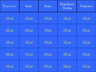
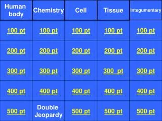
Heart Anatomy Blood Flow CCS ECG Heartium 100 pt 100 pt 100 pt 100 pt 100 pt 200 pt 200 pt 200pt 200 pt 200 pt 300 pt 300 pt 300 pt 300 pt 300 pt 400 pt 400 pt 400 pt 400 pt 400 pt 500 pt 500 pt 500 pt 500 pt 500 pt
Atria: collection chambersseparated by interatrial septumVentricles: discharging chambersseparated by interventricular septumwalls consists of papilary muscleslarger in size and more muscular
1. Atrioventricular valve: Right AVaka tricuspid2. Atrioventricular : Left AV aka bicuspid3.pulmonary semilunar valve 4. Aortic semilunar valve
A backflow of blood caused by a faulty valve is called a __________.
Pericardium: 2 layered sac surrounding the heart tissue; contains serous fluid • Epicardium: lines the outside of the heart • Endocardium: muscle tissue; inner layer of heart tissue
The angle of the heart within the chest should be between ____ and _____?
Pulmonary veins deposit _______ blood into the _______ _________.
This ascending and descending vessel takes blood from the left ventricle to the rest of the body.
DAILY DOUBLE • Describe the flow of blood through the heart? (limited time)
Superior and inferior vena cava-right atrium-tricuspid valve-right ventricle-pulmonary semi-lunar valve-pulmonary arteries-lungs-pulmonary veins-left atrium-mitral valve-left ventricle-aortic valve-aorta-branches of aorta (brachiocephalic, supclavian, carotid)
This part of the CCS is known as the primary pace maker.Where is it located?
SA nodelocated in wall of right atrium near opening of the superior vena cava
The AV node, located in right atrium, send electrical impulses to this bundle of fibers.
These fibers function to contract the ventricles and are stimulated by the ______________.
This node can be called a secondary pacemaker, making the heart beat ~60 beats/min.
The heart muscle is considered ______ ___________ even in the absence of an externally applied nervous impulse. • It can be placed in these certain aqueous solutions to stimulate contractions.
This wave depicted on an ECG involves the depolarization of the ventricular walls.
Repolarization of the ventricles; relaxation of ventricular walls
Which wave on the ECG represents the repolarization of the atria?.
Not depicted on the graph b/c it is masked by the QRS complex, not recorded as a distinct wave.
Cardiac output is equivalent to the heart rate times the amount of blood with each beat
a. What is an arrhythmia? b. List 3 factors that can affect heart rate
Altered heart rhythms or irregular heart beatfactors: temperatureionsdrugsphysical activity (exercise)
Abnormally fast heart rate (100beats/min)caused by shock, drugs, hormones, heart disease, increase in body temperature, exercise, anemia
Justify why ventricular fibrillation is more life threatening than atrial fibrillation.
Ventricles pump O2 blood to systemic and pulmonary circuits…..
Systole involves the _____ of blood through the systemic and ______ circuits.The pressure in the arteries become _______ and the _______ close.