Download
1 / 63
710 likes | 1k Views
ANEMIA. Anemia – Overview. Approach to the anemic patient Definition Manifestations, symptoms, and signs Causes Acute management Peripheral smears Transfusions Indications and when to transfuse What to transfuse Ordering and documentation Transfusion reactions. …so let’s get going.

E N D
Anemia – Overview • Approach to the anemic patient • Definition • Manifestations, symptoms, and signs • Causes • Acute management • Peripheral smears • Transfusions • Indications and when to transfuse • What to transfuse • Ordering and documentation • Transfusion reactions
Definition • What values define anemia? • Values more than 2 standard deviations below the mean • Hgb < 13.5 g/dL or Hct < 41% in men • Hgb < 12.0 g/dL or Hct < 36% in women • Don’t forget…Hgb is a concentration! • May be modified by its content • May be modified by its dilutent (plasma) • Assess the patient’s volume status! • Hemoconcentrated – dehydration • Hemodiluted – after receiving significant IVF • Falsely appearing normal – acute hemorrhage
Assess the patient • History, Physical, Laboratory evaluation • Stable vs. Unstable • Acute anemia due to hemorrhage • Signs/Symptoms of intravascular volume depletion • Pallor • Diaphoresis • Tachypnea • Cold, clammy extremities • Hypotension • Tachycardia • Shock • Anemia that develops slowly • May be recent, subacute, or lifelong • Not usually accompanied by signs of intravascular volume depletion, and symptoms subtle
Symptoms Fatigue Dyspnea Palpitations Worsening angina symptoms or claudication GI disturbances – anorexia, nausea, bowel irregularity Abnormal menstrual patterns Signs Pallor Tachypnea Tachycardia Wide pulse pressure Hyperdynamic precordium Jaundice or splenomegaly Common Symptoms and Signs
Physical Exam • Other Clues • Glossitis – iron, folate, B12 deficiency • Jaundice or Scleral icterus – hemolysis • Splenomegaly – severe thalassemia, chronic hemolytic anemia, leukemia/lymphoma, myeloproliferative syndrome • Neurologic Abnormalities - paresthesias/ataxia B12 deficiency
Causes of Anemia • Kinetic Approach • Blood loss • Inadequate production of RBCs • Hemolysis
Obvious bleeding Trauma Melena Hematemesis Menometorrhagia Occult bleeding Slowly bleeding ulcer or carcinoma Induced bleeding Repeated lab testing Hemodialysis losses Post-surgical bleeding Retroperitoneal space Upper thigh Blood Loss • Most common cause of anemia
Blood Loss • Iron Deficiency • With loss of RBCs, loss of iron will eventually lead to iron deficiency anemia • Iron Stores Depleted • 1200 mL of blood loss – Males • 600 mL of blood loss – Females • 25% of menstruating females have no iron stores • Any amount of blood loss could result in anemia
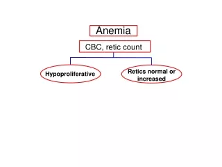
Decreased RBC Production • Rate of production < rate of destruction • Reticulocyte count < 2% • Three morphologies • Microcytic (MCV < 80) • Normocytic (MCV 80 – 99) • Macrocytic (MCV ≥ 100)
Decreased RBC Production: Microcytic • Iron deficiency • Anemia of chronic disease • Copper deficiency • Lead poisoning • Congenital or acquired sideroblastic anemia
Decreased RBC Production: Normocytic • Early B12 or folate deficiency • Dietary loss • Pernicious anemia, sprue, iron deficiency • Anemia of chronic disease • Infection (ie. TB), Inflammation, Malignancy • Bone marrow suppression • Aplastic anemia, acquired red cell aplasia, irradiation • Chronic Renal Insufficiency • Endocrine • hypothyroidism, hypopituitarism • Meds • sulfa drugs, penicillins, anti-epileptics
Decreased RBC Production: Macrocytic • B12 or folate deficiency • Myelodysplastic syndrome • Aplastic Anemia or Pure Red Cell Aplasia • MEDS: Hydroxyurea, zidovudine, ARA-C, methotrexate, azathioprine, 6-MP • Hypothyroidism • EtOH abuse • Multiple Myeloma or other plasma cell disorders • Liver disease / Cirrhosis
Increased RBC Destruction • Hemolysis • RBC life span < 100 days • Occurs when bone marrow cannot keep up with replacement of 5% RBC mass per day • RBC survival ~ 20 days • Reticulocyte count > 3% • Elevated indirect bilirubin • Elevated LDH • Decreased haptoglobin
Inherited Hereditary spherocytosis Sickle cell disease Thalassemia major Antibodies Viruses Drugs AI connective tissue diseases cancer (leukemia/lymphoma) AIHA Acquired Coombs’ positive AIHA TTP/HUS DIC MAHA PNH Malaria Prosthetic heart valves Trauma Hypersplenism Liver disease Increased RBC Destruction
Management • Admitting patients overnight? • What happens when you’re on call? • The middle of the night phone call… • Think of differential diagnoses on your way • Address for most serious life-threatening possibility first • Bedside assessment of patient • Rapid visual assessment of patient’s condition • Vitals • Selective H&P, chart review • Management • Document
Why would they call you for anemia? • “Doctor, this patient is bleeding from…” • “Doctor, this patient’s H/H came back and it’s 6 and 18…” • What do you do?
Management: Hypovolemic patients • Notify resident • Two large-bore IVs (16 gauge) • Type and crossmatch for packed RBCs • May take up to an hour • IV fluids (crystalloid) • Normal Saline or Lactated Ringers • Wide open if in shock • 500 mL to 1000 mL NS bolus if mild/moderate volume depletion • Serial exams are important! for volume status and cardiac exam • Be careful not to put patient in pulmonary edema
Management: Hypovolemic patients • Determine site of hemorrhage • Look for obvious signs of bleeding • IV sites, skin lesions, hematemesis, menstruation • Occult blood loss? • Rectal exam for melena • Flank swelling, flank or periumbilical ecchymosis post-op • Pelvic exam in reproductive age women • Ruptured aneurysm? • Review chart – PMH, meds, recent labs • Surgical consult when appropriate
Management: Normovolemic patients • Exclude active hemorrhage • If Hgb <10 g/dL, consider repeat measurement to exclude lab error • Even if Hgb 10-12 g/dL and normal vitals patients may become unstable quickly if bleeding • What is the patient’s baseline? • Check graphic in Easy-CHCS • If Hgb is more than 0.1-0.2 g/dL less than baseline, assume underlying cause has worsened or secondary factor • Has chronic anemia been evaluated in the past?
Management: Normovolemic patients • Patient is comfortable, normal cardiovascular exam, no suspicion of bleeding… • Baseline studies for further evaluation • MCV (microcytic, normocytic, macrocytic) • RDW - representation of anisocytosis • Reticulocyte count • Peripheral smear
Laboratory Evaluation • Reticulocyte count • High (>3-5) • Increased erythropoietic response to continued hemolysis or blood loss • Low (<2) • Deficient production of RBCs (reduced marrow response) • Concurrent disorder that impairs production (infxn, prior chemotherapy)
Peripheral Smear • Normal peripheral smear • Normal RBC diameter ~ 8-9 microns • About the size of a nucleus of a small lymphocyte • Central pallor ~ 1/3 the RBC diameter
Peripheral Smear • Polychromatic macrocytes • Young reticulocytes with RNA and ribosomes • Slightly larger than other RBCs by 1-2m • Grayish-blue in color • Lacks normal biconcave shape • Represent shift out of marrow into circulation • Can be used to estimate adequacy of erythropoietin response to anemia
He who would cross the Bridge of Death Must answer me These questions three Ere the other side he see….
Peripheral Smear ? • Microcytic anemia – Two examples • Microcytic/Hypochromic • Prominent target cells • Marked deficit of Hgb – flattened appearance • Microcytic/Hypochromic • Pronounced aniso- and poikilocytosis • Presence of cigar-shaped RBCs • Absence of target cells
Macrocytosis Larger than normal RBCs Often oval shaped and well hemoglobinized Hypersegmented neutrophils Larger than normal neutrophils Five or more nuclear lobes Two commonly seen types of anemia Peripheral Smear ?
Peripheral Smear ? • Myelofibrosis - When would you see this • Tear drop cells • Nucleated RBCs • Leukoerythroblastic smear • Seen with immature WBCs
Peripheral Smear • Hemolytic anemia • Inerent defects • Membrane structure, Hgb stability, metabolic fxn • Environmental • trauma, infection, autoimmune • Microspherocytes • Bite Cells • Fragmented RBCs • Abnormal inclusion bodies • Can you name 2 types of inclusion bodies ?
Peripheral Smear • Membrane defects • Uniform elliptical shape • Normal cell indices • Caused by deformation through capillary beds • Cytoskeletal abnormality that prevents recovery of normal shape • Small, dense RBCs • Lack central pallor of normal RBCs • Lack of cell water Higher MCHC • Hereditary spherocytosis • Autoimmune hemolitic anemia
Blood Transfusions • Decision making in a bleeding patient… • Rapid, acute hemorrhage • EBL > 30 – 40%, symptomatic • EBL < 25 – 30%, no uncontrolled hemorrhage • When do patients need PRBCs? • Hgb < 7 g/dL ? • Hgb < 10 g/dL?
Rapid, acute hemorrhage EBL > 30 – 40%, symptomatic EBL < 25 – 30%, no uncontrolled hemorrhage Transfuse PRBC May require uncrossmatched or type-specific blood Transfuse PRBC May require uncrossmatched or type-specific blood Crystalloid/Colloid resuscitation Proceed to PRBC for recurrent signs of hypovolemia Blood Transfusions
Blood Transfusions • What is your transfusion threshold? • How about some help from EBM? • Hebert, et al. (NEJM 1999) looked at critically ill euvolemic patients • 418 transfused for Hgb < 7 g/dL, 420 transfused for Hgb < 10 g/dL • 30-day mortality was similar • These results suggest transfusing at Hgb < 7 g/dL is at least as effective as, and possibly superior to, transfusing at Hgb < 10 g/dL in critically ill patients • Possible exception for patients with AMI and UA • Hebert, et al. (Crit Care Med 2001) did a multi-center trial in critically ill pts with CV disease • 357 pts were randomly assigned to be transfused for Hgb < 7 or < 10 g/dL • There was no differences in overall mortality for the entire group • There was a higher mortality in the subgroup of 257 patients with severe ischemic heart disease who were transfused for Hgb < 7 g/dL (not statistically significant) • Rao, et al. (JAMA 2004) – retrospective study of transfusion in pts ≥ 65 with AMI • 30-day mortality reduced when pts received transfusions for Hct ≤ 30 percent • Possible effectiveness in pts with a Hct ≤ 33 percent
Blood Transfusions • What about transfusing… • Platelets • < 50,000 if acutely bleeding • < 20,000 if not bleeding and febrile • < 10,000 if not bleeding and afebrile • Cryoprecipitate • If patient is in DIC and fibrinogen < 100 • Some say 175 if acutely bleeding • FFP • Emergency reversal of warfarin therapy in bleeding patient • Factor replacement in DIC
Blood Transfusions • What to transfuse and expected increase • PRBC’s: • 1 unit raises Hct by 3 – 4% and Hgb by about 1 gm/dl • unless there is continued bleeding • Platelets: • 1 “six pack” should raise the platelet count by 25,000/mcL • FFP: • Prepared from a single unit of whole blood, contains all coagulation factors and proteins in 1 unit of blood • Cryoprecipitate: • Concentrated preparation of FFP containing only Factors VIII (100 units), XIII, fibrinogen (200 mg) and vWF – final volume is 10-15 mL • Ten bags of cryo (2g fibrinogen) will raise fibrinogen level about 70 mg/dL
Blood Transfusions • Compatability testing • Fully crossmatched RBCs (T&C) • Patients requiring elective transfusion of RBCs • Type, antibody screen, major crossmatch of each unit • Indirect antiglobulin test for minor blood group antibodies • Tests donor cells against patient serum for ABO compatibility • May take several hours • Type and Screen (T&S) • Surgical procedures when blood is occasionally needed • ABO and Rh type • Serum tested for RBC antibodies • If none, typed blood set aside, but not crossmatched • If patient requires transfusion, crossmatched RBC can be made available quickly • If emergency, type-specific blood transfused, crossmatch after
Blood Transfusions • Compatability testing • Emergency crossmatch • Urgently needed blood • Type and crossmatch of RBCs in 15-30 min once pt’s blood is received • If pt has already been transfused, they usually have a sample already • Uncrossmatched/type-specific RBCs • If pt cannot wait 15-30 min for crossmatched or type-specific blood or if pt’s blood type is unknown • Type O-(Rh)negative • Once O-negative is exhausted, O-positive may be substituted with little risk • Conversion to type-specific, crossmatched blood should occur as soon as possible • Risk of inducing antibody only important in women of childbearing age
Blood Transfusions • Leukoreduced RBCs • HLA alloimmunization against class I antigens does not occur • Leukoreduced RBCs do not transmit CMV • Indicated for chronically transfused patients, potential transplant recipients, patients with previous febrile nonhemolytic reactions and CMV negative patients at risk • Irradiated RBCs • Eliminates immunologically competent lymphocytes • Prevents the occurrence of GVHD in patients who have hereditary immune deficiency states • Washed RBCs • Should be considered in patients in whom plasma proteins may cause a serious reaction • IgA deficiency, anaphylaxis with previous transfusions
ANEMIA Document, document, document! You must write a note regarding the indication of transfusion or at least comment on it in your daily progress note X X X The Black Knight 20/123-45-6789
KJ KJ WARD STAFF KJ KJ KJ The Black Knight 20/123-45-6789
Transfusion ReactionsAcute Hemolytic Transfusion Reaction • Due to ABO incompatibility • Incidence 1:250,000-1:1,000,000 with mortality 17-60% • Sx include: fever, chills, vomiting, flank pain, dyspnea, hypotension, tachycardia • Pink plasma and red urine due to intravascular hemolysis • May lead to DIC, shock, and acute renal failure due to ATN
Transfusion ReactionsDelayed Hemolytic Transfusion Reaction • Caused by anamnestic antibody response to specific transfused RBC antigens previously encountered by transfusion, transplantation, or pregnancy • Clinical presentation • Usually asymptomatic • Hct • slight fever • unconjugated bilirubin • spherocytosis on peripheral smear • Hemolysis is usually extravascular, gradual, and less severe than with acute reactions • Rapid hemolysis can occur • Incidence 1:1000
Transfusion ReactionsFebrile nonhemolytic transfusion reaction • Most common transfusion reaction • Occur within 1 – 6 hrs after transfusion RBCs or platelets • Fever, with or without chills or dyspnea • Sx may mimic AHTR or infection – don’t ignore • Incidence: 1% of PRBC transfusions and 30% of platelet transfusions
Transfusion ReactionsTransfusion-related Acute Lung Injury • TRALI is a pulmonary agglutinin reaction • Pathogenesis – 2 stimuli needed • primes neutrophils and activates endothelial cells, leading to increased expression of adhesion molecules • Ex. recent surgery, cytokine administration, massive blood transfusion, or active infection • activates neutrophils causing release of toxic mediators, endothelial damage, and increased capillary permeability • lipid-soluble species that is formed during the storage of banked blood • Causes acute respiratory distress, hypoxemia, hypotension, fever, and pulmonary edema, initially without signs of left ventricular failure • Occurs within 2 – 4 hrs of beginning the transfusion • Incidence 1:2000 • More favorable prognosis than ARDS • Mortality in ≤ 10% of cases of TRALI • Recovery is generally complete within 96 hrs of onset