PAPILLOEDEMA
420 likes | 2.74k Views
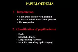
PAPILLOEDEMA. 1. Introduction. Circulation of cerebrospinal fluid Causes of raised intracranial pressure Hydrocephalus. 2. Classification of papilloedema. Early Established (acute) Longstanding (chronic) Atrophic (secondary optic atrophy).

PAPILLOEDEMA
E N D
Presentation Transcript
PAPILLOEDEMA 1. Introduction • Circulation of cerebrospinal fluid • Causes of raised intracranial pressure • Hydrocephalus 2. Classification of papilloedema • Early • Established (acute) • Longstanding (chronic) • Atrophic (secondary optic atrophy)
Circulation of cerebrospinal fluid a (a) Subarachnoid space b (b) Lateral ventricle c d (c) Third ventricle e (d) Aqueduct (e) Fourth ventricle
Causes of Raised Intracranial Pressure 1. Space-occupying lesions 2. Blockage of ventricular system 3. Obstruction of CSF absorption 4. Benign intracranial hypertension (pseudotumour cerebri) 5. Diffuse cerebral oedema 6. Hypersecretion of CSF
Hydrocephalus Dilated cerebral ventricles Communicating - obstruction to CSF flow in basilar cisterns or cerebral subarachnoid space Non-communicating - obstruction to CSF flow in ventricular system or at exit of foramina of fourth ventricle
Early papilloedema • VA - normal • Mild disc hyperaemia • Indistinct disc margins - initially nasal • Mild venous engorgement • Normal optic cup • Spontaneous venous pulsation - absent (also absent in 20% of normals)
Established papilloedema (acute) • VA - usually normal • Severe disc elevation and hyperaemia • Very indistinct disc margins • Obscuration of small vessels on disc • Marked venous engorgement • Reduced or absent optic cup • Haemorrhages + cotton-wool spots • Macular star
Longstanding papilloedema (chronic) • VA - variable • Marked disc elevation but less hyperaemia • Disc margins - indistinct • Variable venous engorgement • Absent optic cup
Atrophic papilloedema (secondary optic atrophy) • VA - severely decreased • Mild disc elevation • Indistinct disc margins • Disc pallor with few crossing vessels • Absent optic cup