Download
1 / 30
320 likes | 638 Views
Visual Function in Friedreich Ataxia. Laura J. Balcer, M.D., M.S.C.E. University of Pennsylvania. Reasons for Understanding Visual System in FA. Importance of vision in controlling ataxia Ease and quantification of measurement of change. Visual Function in Friedreich Ataxia.

E N D
Visual Function in Friedreich Ataxia Laura J. Balcer, M.D., M.S.C.E. University of Pennsylvania
Reasons for Understanding Visual System in FA • Importance of vision in controlling ataxia • Ease and quantification of measurement of change
Visual Function in Friedreich Ataxia Two components to vision: Efferent function: how the eyes move Afferent function: how eyes + brain see
Wiring Diagram of Eye Movements in Ataxia
Efferent Visual Function in Friedreich Ataxia Square-wave jerks: What are they? Fast, random oscillations of both eyes Localize to cerebellum
Square-Wave Jerks Do they matter? Rarely lead to symptoms Worsening of low-contrast letter acuity? No need to treat Can treat oscillopsia with lioresal
Afferent Visual Function in Friedreich Ataxia Afferent function How you see Input to both eyes project back to brain through optic nerve Synapses in lateral geniculate nucleus, project to cortex
Eyes-light rays focused, converted into neural impulses Optic nerve: Impulses transmitted to brain Lateral geniculate: Information sorted Visual cortex: Information decoded
Retinal Nerve Fiber Layer • Light passes to back of eye • Sent out through collected group of axons called optic nerve RNFL = ganglion cell axons
Superior colliculus Optic chiasm Anatomy Cerebral aqueduct Red nucleus Substantia nigra Cerebral peduncle • Optic: • Tract • Chiasm • Nerve Mammillary body
Visual Cortex Higher level visual cortex Primary visual cortex Calcarine sulcus
Visual Function in Friedreich Ataxia Afferent function How you see Input to both eyes project back to brain through optic nerve Synapses in lateral geniculate nucleus, project to cortex
Optic Neuropathy in FA Loss of optic nerve (clinical in 5% , sub-clinical in almost all patients) More common with long GAA repeats, point mutations Usually slowly progressive Can be precipitous Modest spontaneous recovery occasionally observed
Symptoms of Optic Neuropathy in FA Decreased visual acuity: may be subtle Loss of color vision Loss of low contrast vision
Evaluation of Optic Neuropathy in FA Look for other causes- B12 deficiency Glaucoma Vitamin E deficiency Visual Fields MRI if asymmetric, other question
Visual Function in Friedreich Ataxia Lateral geniculate cell loss Similar effect to optic nerve loss
Central Nervous System Visual Axonal Loss • Abnormal tensor MRI signal in visual axon tracts (white matter) • Significance unknown
Approaches to Vision in FA: What’s a Patient to Do? • See ophthalmologist yearly • Get best refraction (glasses, contacts) • Use proper lighting • Posture stabilization • Diabetic control
Reasons for Understanding Visual System in FA Importance of vision in controlling ataxia Ease and quantification of measurement of change
Visual Pathways as a Model for Testing FA Therapies • Sensitive visual function tests • Structure-function correlation captured by ocular imaging
Neurologic Outcomes: FA Composite Test Multidimensional: Timed 25-foot walk (T25W) Nine hole peg test (9HPT) Low contrast letter acuity Correlates with ADL, quality of life, neurologic exam Increasing use as FA trial outcome
Standardized ETDRS format More sensitive than visual acuity, contrast sensitivity Worsening by 2 lines = clinically significant change Scores demonstrate treatment effects in trials and correlate with MRI (in MS), QOL Measuring Visual Function: Low-Contrast Letter Acuity
Does Low-Contrast Acuity Reflect Quality of Life?...Yes! * 25-Item National Eye Institute Visual Function Questionnaire Mowry et al., J Neurol Neurosurg Psychiatry, 2008.
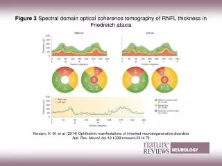
Retinal Nerve Fiber Layer (RNFL) Imaging • Noninvasive, scans take ~15 seconds • Near-infrared light (820 nm) • Reproducible between raters, centers RNFL = ganglion cell axons (non-myelinated)
Retinal Nerve Fiber Layer Thickness Measurement GDx OCT Interference patterns of near infrared light, similar to ultrasound Microtubule birefringence = axonal integrity separate from thickness ** OCT measurements similar to histology ± 5 microns
Low-contrast Acuity RNFL Thickness Low-Contrast Acuity, Optical Coherence Tomography (OCT)
Ultra-High Speed, High-Resolution OCT • Spectral/ Fourier-domain detection • 24,000 A-scans per second • Axial resolution 3.4 µm • 3D images, en face fundus images • Cirrus HD-OCT (Zeiss), RTVue-100 (Optovue), to name a few…
Retinal Nerve Fiber Layer • Light passes to back of eye • Sent out through collected group of axons called optic nerve RNFL = ganglion cell axons
Ganglion Cell Layer Above courtesy of James Fujimoto, Ph.D. High-Speed, Ultra-High Resolution OCT OCT-3 (Stratus)
Conclusions • Eye movement issues in FA are common but of little clinical significance • Afferent vision affected, important • Vision easily quantified, crucial tool for research