Download
1 / 86
870 likes | 951 Views
Learn how to identify and manage neurological emergencies like stroke, seizures, head trauma, and more with a complete guide on conducting thorough neuro exams and recognizing critical conditions. Includes essential stabilization measures and anatomical insights on stroke syndromes.

E N D
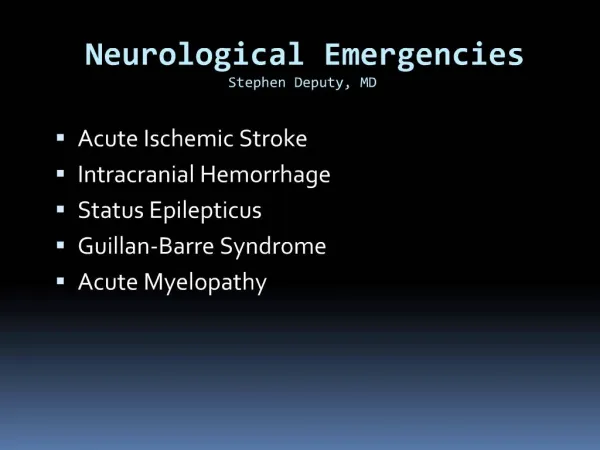
Neurologic Emergency Outline Change in Mental Status / Coma Stroke/TIA Syndromes Seizure & Status Epilepticus Head Trauma Infectious Vertigo/Headaches Peripheral Neuropathies
The Neurologic Exam KEY!! Must do a complete thorough neuro exam to properly identify and diagnose any neurologic abnormality. Exam should include 5 parts: Mental status, level of alertness (GCS) Cranial nerve exam Motor / Sensory exam Reflexes Cerebellar Consider ; MMSE if Psych components
Change in Mental Status / COMA Potential Causes – “AEIOU TIPS” A = Alcohol ( Drugs & Toxins) E = Endocrine, Exocrine, Electrolyte I = Insulin O = Opiates, OD U = Uremia T = Trauma, Temperature I = Infection P = Psychiatric disorder S = Seizure , Stroke, Shock, Space occupying lesion
Change in Mental Status/Coma Temperature Hypothermia: causes coma when Temp<32.0 C Hyperthermia: causes coma when Temp>42.0C Infection Meningitis, Encephalitis, Sepsis Endo/Exocrine, Electrolyte Hypo/Hyperglycemia Hypo/hyperthyroidism Hypo/hypernatremia Hepatic encephalopathy Opiods/ OD / Alcohol Heroin, Psych Meds (TCA’s, SSRI’s)
Change in Mental Status/Coma • T for Trauma
AMS / COMA Physical Exam Pearls Always attempt to get a complete history!! LOOK at your patient! Smell the breath (ketones,alcohol,fetid) Observe respiratory rate & patterns (Cheyne-Stokes) Look for abnormal posturing. Decorticate (Flexion of UE with Extension of LE) Decerebrate (Extension of all Ext.) Look for needle marks, cyanosis, signs of trauma Obtain GCS Score! E4 V5 M 6 If less than 8, IMMEDIATE airway stabilization FIRST priority!!
Glasgow COMA Scale Scores range from 3 (Worst) – 15 (Best) Important for classifying degree of alteration. (Head Trauma) GCS < 8 = INTUBATE!! EYE Opening Response 4 = Spontaneous 3 = To Voice 2 = To Pain 1 = None Remember as “4 eyes”
Glasgow COMA Scale Verbal Response 5 = Oriented and converses 4 = Confused but converses 3 = Inappropriate words 2 = Inappropriate sounds 1= None Remember as “Jackson 5 – sing/voice”
Glasgow COMA Scale Motor 6 = Obeys commands 5 = Localizes pain 4 = Withdraws to pain 3 = Decorticate (flexes to pain) 2 = Decerebrate (extends to pain) 1 = None Remember as “ 6 Cylinder engine – motor”
AMS / COMA Essential Stabilization & Assessment Measures Always assess & stabilize ABC’s first special attention to airway with C-Spine immobilization / protection. Oxygenate! IV line , fluids, Thiamine 100mg IV, 1 amp D 50, & Narcan(if needed) 0.4mg increments until response. Complete history and physical exam after stabilization Radiographic clearance of C-Spine Labs / CT as indicated
Stroke / TIA Syndromes Anatomy of Cerebral Blood Flow Anterior Circulation: 80% of cerebral blood flow originates from the carotids which supplies the Frontoparietal lobes Anterior temporal lobes Optic nerve and retina Posterior Circulation: 20 % of cerebral blood flow which originates from the vertebrobasilar arteries Thalamus & Brainstem Occipital cortex and Cerebellum Upper Spinal cord & Auditory and Vestibular functions in ear Circle of Willis: connects the Anterior and Posterior circulations
Pathophysiology of Stroke / TIA Ischemic Strokes: (thrombi or emboli) Cerebral Thrombi may result from: Atherosclerosis (#1 cause) Infective arteritis Vasculitis Hypercoagulable states Post traumatic carotid or vertebral artery dissections Cerebral emboli may result from: Mural thrombus from heart (#1 cause) Aortic plaques Endocarditis Long bone or Dysbaric injuries (fat / air emboli)
Pathophysiology of Stroke/TIA Hemorrhagic Strokes result from Spontaneous rupture of berry aneurysm or AV malformation (Subarachnoid hemorrhage) Rupture of arteriolar aneurysms secondary to: Hypertension Congenital abnormality Blood dyscrasia / Anticoagulant usage Infection Neoplasm Trauma (Epidural / Subdural Hematomas) Hemorrhagic transformation of embolic stroke
Stroke /TIA Syndromes Type of Stroke (rule of 2/3’s) 2/3 of ALL Strokes are ISCHEMIC 2/3 of these are thrombotic Therefore thrombotic, ischemic strokes most common. Incidence of Stroke Biggest Risk Factors Prior TIA ( 30 % will have stroke in 5 years) HTN Atherosclerosis DM Hyperlipidemia Smoking
Ischemic Stroke Syndromes: Thrombotic vs. Embolic Thrombotic Syndromes Usually slow, progressive onset Sx develop shortly after awakening and are progressive Embolic Syndromes Usually abrupt onset with maximal deficit that tends to improve over time as the embolus breaks up.
Ischemic Stroke Syndromes Middle Cerebral Artery Occlusion (MCA) # 1 type Contralateral hemiplegia, hemianesthesia, and homonymous hemianopsia Upper extremity deficit >> Lower extremity Aphasia (if dominant hemisphere involved) Conjugate gaze impaired in the direction of the lesion
Ischemic Stroke Syndromes Anterior Cerebral Artery Occlusion (ACA) Contralateral leg, arm, paralysis Lower Extremity deficit >> Upper extremity Loss of frontal lobe control Incontinence Primitive grasp and suck reflexes enacted Posterior Cerebral Artery Occlusion (PCA) Ipsilateral CN III palsy, visual loss Contralateral hemiparesis and hemisensory loss Memory loss
Ischemic Stroke Syndromes Vertebrobasilar Artery Occlusion (VBA) Hallmark: Crossed Neurological Deficits CN AND Cerebellar deficits that affect BOTH sides of the body, with contralateral pain and temperature deficits. - Contralateral hemiplegia Ipsilateral CN III palsy with Cerebellar findings. Nausea/Vomiting Vertigo, Nystagmus, Ataxia, Dysarthia Tinnitus, deafness
TIA’s (Transient Ischemic Attacks) Definition: A temporary loss of neurologic function, that resolves completely <24 hours. Clinically; Arm numbness, weakness, HA Facial droop, slurred speech Sx resolved, or improve over time Main point: These patients at high risk for stroke if: >50 HTN, DM, Smoker, Prior TIA in last month Any prior CVA…… ADMISSION IS THE RULE!! Treat as CVA : Head CT (CVA protocol) ASA 81-325mg po Oxygen, 2L NC If cardiac arrythmia (atrial filbrillation) present, consider Heparin ONLY after Head CT and Neuro consultation.
Hemorrhagic Stroke Syndromes (SAH, & Intracerebral) Subarachnoid Hemorrhage Highest incidence in 35-65 year old. Usually from the rupture of a berry aneurysm Clinically: abrupt onset of “worst headache of life” Nuchal rigidity, photophobia, vomiting, retinal hemorrhages. Diagnosis : CT + LP!!!! CT only 92% sensitive within 24 hours of event, loses sensitivity >24 hours out from headache. 72 hours out CANNOT r/o without LP! Management: Consider adding Nimodipine 60 mg Q6 to reduce vasospasm
Hemorrhagic Stroke Syndromes Intracerebral Hypertensive intracerebral hemorrhage MOST common cause. Traumatic, contusion, coup/contracoup Rupture of small blood vessels with bleeding inside the brain parenchyma Putamen Cerebellar Thalamus Pontine ( 3 P’s – pinpoint pontine pupils)
Treatment of Stroke AS ALWAYS – ABC’s FIRST What’s the Serum Glucose?? Consider Thiamine 100mg IV, D 50 bolus if hypoglycemic. Treat Hyperglycemia if Serum Glucose > 300mg/dl Protect the “Penumbra” Keep SBP >90mm Hg Goal keep CPP > 60mm Hg (CPP=MAP-ICP) Treat Fever ( Mild Hypothermia beneficial) Acetaminophen 650mg po or pr, cooling blanket Oxygenate (Keep Sao2 >95%) Elevate head of bed 30 deg. (Clear c-spine) Frequent repeat Neuro checks!! Reassess GCS!
Treatment of Stroke What type of stroke is Present?? Hemorrhagic vs Ischemic Any signs of shift herniation? Neurosurgery evaluation or transfer necessary? Other management adjuncts: Ischemic strokes ASA 81-325mg Patients with Systolic BP >220 , Diastolic>120 need BP control with Nitroprusside or Labetolol. DO NOT OVERTREAT BP or risk extending the infarct. Heparin not shown to be of benefit in recent studies, however, still frequently used. Consult Neurologist before use If used, No bolus, just infusion. Risk of hemorrhagic transformation.
Treatment of Strokes Strokes with Edema, Mass Effect or Shift Load with Phosphenytoin 1000mg for seizure prophylaxis Acute seizure prophylaxis still of benefit. Mannitol, Decadron?? Recently shown to be of NO benefit, some Neurosurgeons still advocate, so consult first. Hyperventilation?? NOT beneficial and perhaps harmful, don’t do it! Thrombolytics??? Ischemic strokes ONLY with large deficit NOT improving. Time from symptom onset <3 hours No ABSOLUTE Contraindications!! Inclusion and Exclusion Criteria Benefit Questionable
Thrombolytic Therapy for Acute Stroke Checklist Answer to ALL must be YES: Age 18 or older Clinical diagnosis of Acute Ischemic Stroke causing a measurable NON improving neurologic deficit. NO high clinical suspicion for SAH Time of onset to treatment is <180 minutes.
Thrombolytic Therapy for Acute Ischemic Stroke Checklist Answer to ALL MUST be NO: Evidence of hemorrhage on CT Active internal bleeding (GI/GU) within last 21 days. Known bleeding diasthesis: Platelets<100,000 Heparin within last 48 hours with elevated PTT Warfarin use with PT > 15 seconds Within 3 months of IC injury, prior surgery or prior ischemic stroke. Within 14 days of serious trauma, major surgery Recent AMI, arterial puncture/LP within 7 days History of prior ICH, AVM, tumor,or aneurysm or seizure at stroke Systolic BP >185mmHg, or Diastolic BP >110Hg
Seizures & Status Epilepticus Background: 1 – 2% of the general population has seizures Primary Idiopathic epilepsy: onset ages 10-20 Secondary Precipitated by one of the following: Intracranial pathology Trauma, Mass, Abscess, Infarct Extracranial Pathology Toxic, metabolic, hypertensive, eclampsia
Seizure Types Generalized Convulsive Seizures (Grand Mal): Tonic , clonic movements, (+) LOC, apnea, incontinence and a post-ictal state Non Convulsive Seizures (Petit Mal) Absence seizures – “blank staring spells” Myoclonic – brief contractions of selected muscle groups Partial Seizures Characterized by presence of auditory or visual hallucinations Simple = somatic complaints + no LOC Complex = somatic complaints + AMS or LOC
Approach for 1st Seizure, New Seizure, or Substance/ Trauma Induced Seizure As always ABC’s First IV, O2, Monitor. Send blood for CBC, Chem 20, Tox screen as appropriate Anticonvulsant levels Prolactin levels / Lactate level CXR / UA/ Head CT Is patient actively seizing? Post ictal? Pseudoseizure? Consider treatment options Complete History and Physical Exam Including detailed Neuro Exam Repeat Neuro evaluations a must!
Guidelines for Postictal Head CT Scans Status Epilepticus ( a true emergency) Abnormal Neuro findings No return to GCS 15 Prolonged HA History of malignancy CHI (Closed Head Injury) HIV infection or high risk for HIV Anticoagulant use Age > 40
Approach to Breakthrough Seizure As Before, But History, History, History!! Main causes of Breakthrough Seizure: Noncompliance with anticonvulsant regimen Start of new medication (level alteration) Antibiotics, OCP’s Infection Fever Changes in body habitus, eating patterns Supratherapeutic level
Status Epilepticus Definition: operationally defined as seizure lasting greater than 5 minutes OR two seizures between which there is incomplete recovery of consciousness. Treatment algorhythm: As before ABC’s IV, O2, Monitor Consider ALL potential causes INH (pyridoxime/B-6 deficiency) Eclampsia Alcoholic (thiamine/B-1 deficiency) Other Tox ingestion (TCA’s, sulfonylurea OD) Trauma
Status Epilepticus Treatment FIRST LINE TREATMENT Lorazepam (Ativan) 2mg/min IV up to 10 mg max. OR Diazepam(Valium) 5mg/min IV or PR up to 20mg SECOND LINE TREATMENT Phenytoin or Fosphenytoin (Cerebyx): 20mg/kg IV at rate of 50mg/min THIRD LINE TREATMENT Get Ready to intubate at this point!! Phenobarbitol 10-20mg/kg @ 60 mg/min
Status Epilepticus Treatment FINAL TREATMENT Barbiturate Coma Pentobarbitol 5mg/kg @ 25 mg/min Stat Neurology consult for evaluation and EEG Pentobarbitol titrated to EEG response. Always get a through HISTORY Possible trauma Medications in house Others sick, symptomatic Overall appearance of patient
Status Epilepticus Adjunctive Treatment by History Thiamine 100mg IV, 1-2 amps D 50 If suspect alcoholic, malnourished, hypoglycemia Magnesium Sulfate 20cc of 10% solution As above of if eclampsia (BP does NOT have to be 200/120!!) Pyridoxine 5 gms IV INH or B-6 deficiency
Closed Head Injury Definitions : Concussion: refers to a transient LOC following head injury. Often associated with retrograde amnesia that also improves. “Coup” = injury beneath the site of trauma “Countrecoup” = injury to the side polar opposite to the traumatized area. Diffuse Axonal Injury : tearing and shearing of nerve fibers at the time of impact secondary to rapid acceleration/deceleration forces. Causes prolonged coma, injury, with normal initial head CT and poor outcome.
Closed head Injury Facts The single most important factor in the neurologic assessment of the head injured patient is level of consciousness. (LOC) Always assume multiple injuries with serious mechanism. ESPECIALLY C - SPINE!!!! Unless hypotensive WITH bradycardia and WARM extremities (spinal cord injury); hypotension is ALWAYS secondary to hypovolemia from blood loss in the trauma patient! The most common intracranial bleed in CHI is subarachnoid hemorrhage.
Closed Head Injuries with Hemorrhage Cerebral Contusion Focal hemorrhage and edema under the site of impact. Susceptible areas are those in which the gyri are in close contact with the skull Frontal lobe Temporal lobes Diagnostic Test of Choice: NC Head CT Treatment: Supportive with measures to keep ICP normal. Repeat Neuro checks. Repeat Head Ct in 24 hours. Good prognosis.
Subdural Hematoma Occurs secondary to acceleration/decelleration injury with resultant tearing of the bridging veins that extend from the subarachnoid space to the dural sinuses. Blood dissects over the cerebral cortex and collects under the dura overlying the brain. Patients at risk: Alcoholics Elderly Anticoagulant users Appears as “sickle shape” and does not extend across the midline
Epidural Hematoma Occurs from blunt trauma to head especially over the parietal/temporal area. Presents as LOC which then patient has lucid interval then progressive deterioration, coma , death. ( Patient talks to you & dies!) Commonly associated with linear skull fracture Mechanism of bleed is due to tear of artery, usually middle meningeal. PE reveals ipsilateral pupillary dilitation with contralateral hemiparesis. CT Scan : a BICONVEX (lens) density which can extend across the midline
Management of Closed Head Injuries As always ABC’s with C-Spine precautions IV, O2, Monitor. Stabilize and resuscitate Sao2>95% SBP>90 Treat Fever Head of Bed 30% (once C-Spine cleared) Stat Head CT with Stat Neurosurgical evaluation for surgical lesions. Repeat Exams, looking for signs of herniation.
Signs of Herniation / Increased ICP Headache, nausea, vomiting Decreasing LOC Sixth nerve paresis (one or both eyes adducted) Decreased respiratory rate Cushing reflex (hypertension/bradycardia/bradynpea) Papilledema Development of signs of herniation Fixed and dilated pupil Contralateral hemiparesis Posturing
Herniation Syndromes CPP = MAP – ICP: Must keep CPP >60 mm Hg Uncal Herniation: Occurs when unilateral mass pushes the uncus (temporal lobe) through the tentorial incisa, presenting as: Ipsilateral pupil dilatation Contralateral hemiparesis Deepening coma Decorticate posturing Apnea and death