Download

1 / 86
860 likes | 884 Views
This article discusses the fractures of the pelvis and acetabulum in pediatric patients, including the anatomical and developmental aspects, as well as the examination and evaluation methods.

E N D
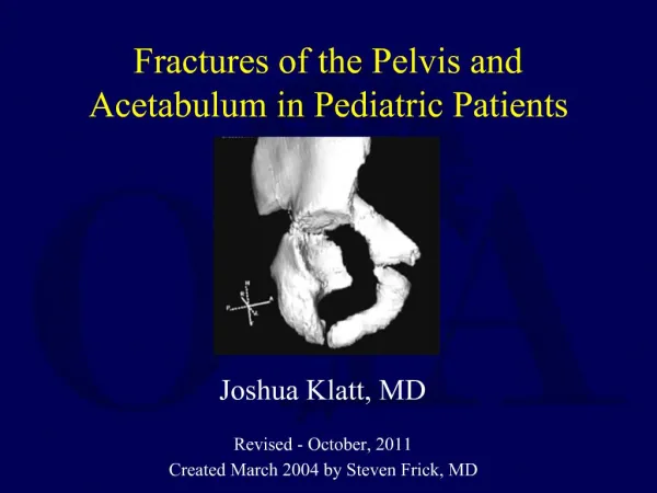
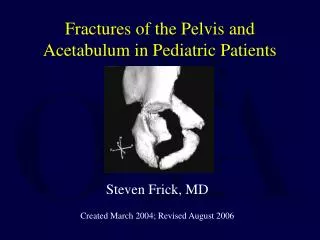
Fractures of the Pelvis and Acetabulum in Pediatric Patients Joshua Klatt, MD Revised - October, 2011 Created March 2004 by Steven Frick, MD
The Child’s Pelvis • Fundamental differences: • Bones are more malleable • Cartilage is capable of absorbing more energy • SI joint and symphysis are more elastic • Triradiate Cartilage • Injury causing growth arrest may lead to significant deformity Schlickwei W, Keck T. Pelvic and acetabular fractures in childhood. Injury. 2005; 36(suppl 1):A57-A63.
Elasticity of Joints • Sacroiliac joint and pubic symphysis are more elastic • Allows significant displacement before fx • Allows for single break in the ring • Thick periosteum • Apparent dislocations may have a periosteal tube that heals like a fracture Schlickwei W, Keck T. Pelvic and acetabular fractures in childhood. Injury. 2005; 36(suppl 1):A57-A63.
Pelvic Anatomy • 3 primary ossification centers: • Ilium – appears at 9 wks • Ischium – appears at 16 wks • Pubis – appears at ~20 wks • Endochonral ossification, just like long bones Delaere O, Dhem A. Prenatal development of the human pelvis and acetabulum. Acta Orthop Belg. 1999;65(3):255-60.
Acetabular Anatomy • The 3 distinct physes of each bone making up the triradiate cartilage allow hemispheric growth of both the acetabulum and pelvis. • The 3 ossification centers meet and fuse at the triradiate cartilage at age 13-16 years Ponseti, I. Growth and development of the acetabulum in the normal child. Anatomical, histological, and roentgenographic studies. J Bone Joint Surg Am. 1978;60(5):575-85.
Triradiate Cartilage Complex • Separates the ilium, the pubis and the ischium Ponseti, I. J Bone Joint Surg Am. 1978.
Infant Acetabulum Histologic section of infant acetabulum • Acetabular cartilage • Triradiate cartilage • Labrum • Pulvinar • Capsule • Ilium Ponseti, I. J Bone Joint Surg Am. 1978.
Development of the Acetabulum • Interstitial growth within the horizontal flange of the triradiate cartilage contributes to the normal growth of the distal third of the ilium. • Enlargement of the acetabulum during growth is likely the result of interstitial growth within the triradiate cartilage. Ponseti, I. J Bone Joint Surg Am. 1978.
Development of the Acetabulum • Development of concavity is a response to pressure from the femoral head • In DDH with a dislocation the acetabulum will not develop appropriately • Depth of the acetabulum results from: • Interstitial growth in the acetabular cartilage • Appositional growth of the periphery of this cartilage • Periosteal new bone formation at the acetabular margin.
Puberty • 3 secondary ossification centers appear in the hyaline cartilage of the acetabulum • Os acetabuli • Epiphysis of the pubis • Forms most of anterior wall • Acetabular epiphysis • Epiphysis of the ilium • Forms most of superior acetabulum • Secondary ossification center of the ischium • Forms much of posterior wall
Secondary Ossification Centers • OA - Os Acetabuli • AE - Acetabular Epiphysis • PB - Pubic Bone • SCI – Secondary ossification center of ischium • Ossification centers appear at age 8 to 9 yrs and fuse around 17 – 18 yrs SCI Ponseti, I. J Bone Joint Surg Am. 1978.
Anatomy • Other secondary ossification centers of the pelvis • Iliac crest • Ischial apophysis • Anterior inferior iliac spine • Pubic tubercle • Angle of the pubis • Ischial spine • Lateral wing of the sacrum
Secondary Ossification Center • Iliac Crest : first seen at age 13 to 15 and fuses at age 15 to 17 years • Used in Risser staging • Ischium : first seen at age 15 to 17 and fuses at age 19 to 25 years • ASIS : first seen about age 14 and fusing at age 16 *Important to know these secondary ossification centers so they will not be confused with avulsion fractures
Weakness of Cartilage • Avulsion fractures occur more often in children and adolescents through an apophysis • Fractures of the acetabulum into the triradiate cartilage may occur with less energy than adult acetabular fractures
History and Associated Injuries • Pelvic ring and acetabular fractures usually involve high energy injuries • Associated injuries • Orthopaedic – long bone or spine fractures • Urologic – bladder rupture • Vascular – less frequent than in adults, rarely life threatening
Physical Examination • A, B, C’s • Trauma evaluation • Orthopaedic exam of extremities and spine • Systematic approach to the pelvis
Examination of the Pelvis • Areas of contusion, abrasion, laceration, ecchymosis, or hematoma, especially in the perineal and pelvic areas, should be noted • Rule out open fractures in perineum/genital/rectal areas • Palpate landmarks • Anterior superior iliac spine • Crest of the ilium • Sacroiliac joints • Symphysis pubis
Examination of the Pelvis • Neurologic and vascular exam of the lower extremities • Provocative Tests • Compress the pelvic ring with anterior-posterior and lateral compression stress • The range of motion of the extremities, especially of the hip joint, should be determined
Radiographic Evaluation • There is no standard algorithm for which films to obtain in children • AP pelvis • Judet views for acetabular involvement • Inlet/Outlet views for pelvic ring injuries • Computed tomography • 2D and possibly 3D reconstruction • Cystography/urography if blood at meatus or on bladder catheterization
Pelvic Avulsion Fractures • Caused by forceful contraction at sites of muscle attachments through apophyses • Iliac wing – tensor fascia lata • Anterior superior iliac spine – sartorius • Anterior inferior iliac spine – rectus femoris • Ischium – hamstrings • Lesser trochanter - iliopsoas
Relative Percentages of Pelvic Avulsion Fracture Locations • Ischial tuberosity – 54% • AIIS – 22% • ASIS – 19% • Pubic Symphysis – 3% • Iliac Crest – 1% http://crashingpatient.com Rossi F, Dragoni S. Acute Avulsion Fractures of the Pelvis in Adolescent Competitive Athletes. Skeletal Radiol. 2001;30(3):127-31.
Pelvic Ring Injuries • Often high energy mechanism • MVA • Auto-pedestrian • Fall from height • Often other fractures present • Traumatic brain injury (TBI) • Intra-abdominal injuries • Urologic injuries • Neurologic and vascular injuries may occur with severe disruptions Torode I, Zieg D. Pelvic fractures in children. J Pediatr Orthop 1985;5:76-84.
Classification of Pelvic Injuries in ChildrenTorode and Zieg modification of Watts classification • Type I – avulsion fractures • Type II - Iliac wing fractures • Type III – stable pelvic ring injuries • Type IV – any fracture pattern creating a free bony fragment (unstable pelvic ring injuries) Torode I, Zieg D. Pelvic fractures in children. J Pediatr Orthop 1985;5:76-84.
Tile Classification • Applicable in patients near skeletal maturity • More often adult type patterns • Type A – Stable • Type B – Rotationally unstable, vertically stable • Type C – Rotationally and vertically unstable Tile M. Acute Pelvic Fractures: I. Causation and Classification? J Am Acad Orthop Surg. 1996;4(3):143-151.
Treatment Options • Bedrest • Spica cast • Restricted weight bearing • Skeletal traction • External fixation • ORIF
Treatment Differences • Children tolerate bedrest/traction/immobilization better than adults • Pubic symphyseal and SI disruptions may be able to be treated closed because of potential for periosteal healing • Operative fixation should spare growth plates when possible • When not possible consider temporary (4-6 weeks) fixation across physes with smooth pins or early hardware removal Holden C, et al. Pediatric pelvic fractures. J Am Acad Orthop Surg. 2007;15:172-7.
Pelvic Ring Injuries *Often crush mechanism and can have severe soft tissue injuries as well.
Treatment • Most avulsion injuries and Tile A fractures treated with restricted or no weight bearing • Most Tile B fractures treated nonoperatively unless major deformity • Tile C fractures may need stabilization Holden C, et al. Pediatric pelvic fractures. J Am Acad Orthop Surg. 2007;15:172-7.
Treatment Caveats • Treat older children and adolescents with pelvic injuries like adults • In general, pelvic injuries where posterior ring disruptions are displaced or unstable need operative treatment • Only anterior ring may need stabilization • And for shorter time period, if using external fixation Holden C, et al. Pediatric pelvic fractures. J Am Acad Orthop Surg. 2007;15:172-7.
13 year old, bilateral pubic rami fractures with left SI disruptionsubtrochanteric femur fracture
Pediatric Acetabular Fractures • Constitute only 1% to 15% of pelvic fractures in children • Much more common after the triradiate cartilage closes (12 yrs in girls, 14 yrs in boys) • Mechanism of injury similar to that in adults • Force transmitted through femoral head • Position of leg relative to pelvis and location of impact determine fracture pattern
Pediatric Acetabular Fractures • Often associated with hip dislocation • The distribution of types is different than adults • More often transverse than both column • Historically treated nonoperatively • Achieving congruent reduction with closed, conservative treatment is difficult and often impossible • Many think that the role of surgical treatment in children is expanding Heeg M, de Ridder VA. Acetabular fractures in children and adolescents. Clin Orthop Relat Res 2000;376:80–6.
Pediatric Acetabular FracturesClassification • Growth plate injury • Use Salter-Harris classification • Bucholz suggested that there are common injury patterns • Letournel system most frequently used • Same as used for adults • Watts classification also sometimes used Bucholz, et al. Injury to the acetabular triradiate physeal cartilage. J Bone Joint Surg Am 1982;64(4):600-9.
Pediatric Acetabular FracturesClassification • Letournel system • Type A – Single wall or column • Type B – Fractures involving 2 columns • Type C – Fractures involve both columns and separate dome fragment from axial skeleton Judet, et al. Fractures of the acetabulum: classification and surgical approaches for open reduction. J Bone Joint Surg Am 1964;46:1615-46.
Pediatric Acetabular FracturesClassification • Watts classification • Type A – Small fragments that most often occur with hip dislocation • Type B – Stable linear fractures without displacement in association with pelvic fractures • Type C – Linear fractures with hip joint instability • Type D – Fractures secondary to central fracture-dislocation of the hip Watts HG. Fractures of the pelvis in children. Orthop Clin North Am 1976;7:615-624.
Pediatric Acetabular FracturesClassification • Injuries to the triradiate cartilage constitute physeal trauma • Bucholz Classification • Two basic patterns • Shearing Type (Salter-Harris Type I or II) • Crushing or Impaction Type (Type IV) Bucholz, et al. Injury to the acetabular triradiate physeal cartilage. J Bone Joint Surg Am 1982;64(4):600-9.
Bucholz, et al. Injury to the acetabular triradiate physeal cartilage. J Bone Joint Surg Am 1982;64(4):600-9.
Pubic ramus fractures and triradiate cartilage injury • OFTEN associated ring injury • Watts Type B injury • Bucholz Shearing Type • Salter-Harris II
Shearing Type • Blow to the pubic or ischial ramus or the proximal end of the femur • Injury at the interface of the 2 superior arms of the triradiate cartilage and the metaphysis of the ilium • A triangular medial metaphyseal fragment (Thurston-Holland sign) may be seen in Salter-Harris Type II injuries Bucholz, et al. Injury to the acetabular triradiate physeal cartilage. J Bone Joint Surg Am 1982;64(4):600-9.
Shearing Type • Effectively splits the acetabulum into superior (main weight-bearing) one-third and inferior (non-weight-bearing) two-thirds • Germinal zones contained within the physes often unaffected • Favorable prognosis for continued relatively normal growth and development of the acetabulum Bucholz, et al. Injury to the acetabular triradiate physeal cartilage. J Bone Joint Surg Am 1982;64(4):600-9.
Shearing Pattern with Central Protrusio of Femoral Head • Watts Type D injury • Bucholz Shearing Type
Crushing or Impaction Type • Difficult to detect on initial radiographs • Narrowing of the triradiate space suggests this injury pattern (rarely seen) • Premature closure of the triradiate cartilage appears to be the usual outcome • The earlier in life the premature closure occurs, the greater the eventual acetabular deformity Bucholz, et al. Injury to the acetabular triradiate physeal cartilage. J Bone Joint Surg Am 1982;64(4):600-9.
Triradiate Cartilage • Fractures through this physeal cartilage in children can ultimately cause: • Growth arrest • Leg-length discrepancy • Faulty development of the acetabulum Heeg, et al. Injuries of the acetabular triradiate cartilage and sacroiliac joint. J Bone Joint Surg Br 70:34-37,1988.
Age is a significant risk factor in the development of post-traumatic acetabular dysplasia. Children younger than ten years of age at the time of injury are at greatest risk Bucholz, et al. Injury to the acetabular triradiate physeal cartilage. J Bone Joint Surg Am 1982;64(4):600-9.