
ONCOGENES
E N D
Presentation Transcript
M.Prasad Naidu MSc Medical Biochemistry, Ph.D.Research Scholar CANCER
The term cancer applies to a group of diseases in which cells grow abnormally • and form a malignant tumor. • Malignant cells can invade nearby tissues and metastasize (establish secondary • areas of growth). • This aberrant growth pattern results from mutations in genes that regulate proliferation, differentiation, and survival of cells in a multicellular organism. • Because of these genetic changes, cancer cells no longer respond to the signals that govern growth of normal cells
Oncogenes–The genes involved in the development of cancer normal cells do contain DNA sequence similar to viral oncognenes To distinguish these two genes: V-src (viral gene) and C-src (cellular gene) • Protooncogenes– normal constituents of cells whose function is to promote proliferation or cell survival. These genes can code for growth factors, growth factor receptors, signal transduction proteins, intracellular kinases and transcription factors. • Tumor suppressor genes(normal growth suppressor genes) -- encode proteins that inhibit proliferation, promote cell death, or repair DNA Activation of oncogenes or absence /inactivation of tumor suppressor genes can lead to cancer.
Protooncogenes are regulatory genes • Products of many oncogene are polypeptide growth factor ex: sis gene produce PDGF - normal wound healing. • Product act as receptor for growth factor ex: erb-B produces receptor for EGF • Some act on key IC pathway involved in growth control ex: Src Product receptor of EGF, insulin, PDGF. • C-oncogenes are under the control of regulatory genes & expressed only when required. • When virus enters, an extra oncogene is inserted so as to produce continuous expression of gene leading to uncontrolled cellular activity & malignant transformation.
Virus Chemical carcinogens Chromosomal translocation γ-rays Spontaneous mutation All such factors may converge into one biochemical abnormalities “Activation of protooncogenes” leading to malignancy Many factor activate protooncogenes • Because neoplasia is a multistep process, more than one of these mechanisms often contribute to the genesis of human tumors by altering a number of cancer-associated genes. • Full expression of the neoplastic phenotype, including the capacity for metastasis, usually involves a combination of protooncogene activation and inactivation tumor suppressor gene.
5 mechanisms of activation : • Promoter insertion • Enhancer insertion • Chromosomal translocation • Gene amplification • Point mutations
1. Promoter Insertion • Certain retro viruses lack oncogenes ( eg : avian leukemia viruses ) but may cause cancer over a long period of time.
Viral insertion into chromosomes: • In retrovirus, cDNA is made from their RNA by enzyme reverse transcriptase. • cDNA gets inserted into host genome • Integrated dscDNA provirus • This proviral DNA takes over the control of transcription of cellular chromosomal DNA & transforms the cell. eg: Avian leukemia
3. Chromosomal translocation • Rearrangement of genetic material by splitting off a small fragment of chromosome which is joined to another chromosome. • Over expression of proto oncogenes eg: Burkitts lymphoma Chronic myeloid leukemia
The bcr/abl fusion, created on the chromosome 22, encodes a chimeric protein of 210 kDa, with increased tyrosine kinase activity and abnormal cellular localization. 20% of cases of ALL. Overexpression of the bcl-2 protein inhibits apoptosis, leading to an imbalance between lymphocyte proliferation and programmed cell death. • c-myc finds itself in a region of active gene transcription, and it may simply be the overproduction of the c-myc product (a transcription factor essential for cell division) that propels the lymphocyte down the pathway towards cancer.
4. Gene amplification • Certain DNA sequence is amplified several fold in some cancers. • Gene amplification was first discovered as a mechanism by which some tumor cell lines can acquire resistance to growth-inhibiting drugs. • Methotrexate becomes inactive due to gene amplification resulting in a several fold increase in activity of DHR. • Studies then demonstrated that three protooncogene families-myc, erb B, and ras-are amplified in a significant number of human tumors. • About 20% to 30% of breast and ovarian cancers and some types of SCC show c-myc amplification. • Amplification of N-myc correlates strongly with advanced tumor stage in neuroblastoma
5. Mutations: • Mutations activate protooncogenes through structural alterations. These alterations, which usually involve critical protein regulatory regions, often lead to the uncontrolled, continuous activity of the mutated protein. • Various types of mutations, such as base substitutions, deletions, and insertions, are capable of activating protooncogenes. • In human tumors the most characterized oncogene mutations are base substitutions (point mutations) that change a single amino acid within the protein. • Mutations in DNA that give rise to cancer may be inherited or caused by chemical carcinogens, radiation, viruses, and by replication errors that are not repaired.
Point mutation • Point mutations are frequently detected in the ras family of protooncogenes (K-ras, H-ras, and N-ras). • Single most dominant cause of many human tumor. • Ras protein M.W 21000(P21) • Inactive ras is in bound state with GDP. • When cells are stimulated by GF, ras P21 get activated by exchanging GDP for GTP. • In normal cells, the activity of ras P21 is short lived because of GTPase activity. • Point mutation cause altered ras P21 lacking GTPase activity
Studies have found K-ras mutations in about 30% of lung adenocarcinomas, 50% of colon carcinomas, and 90% of carcinomas of the pancreas. • N-ras mutations – hematologic malignancies • Another significant example of activating point mutations is represented by those affecting the ret protooncogene in multiple endocrine neoplasia type 2A syndrome (MEN2A)
Growth factors • The genes for both growth factors and growth factor receptors are oncogenes. • Growth factors generally regulate growth by serving as ligands that bind to cellular receptors located on the plasma membrane (cell-surface receptors) . • Binding of ligands to these receptors stimulates a signal transduction pathway in the cell activating the transcription of certain genes. • If too much of a growth factor or a growth factor receptor is produced, the target cells may respond by proliferating inappropriately. • Growth factors receptors may also become oncogenic through translocation or point mutations.
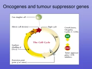
Because the cell is committed to DNA replication and division once it enters the S phase, multiple regulatory proteins are involved in determining whether the cell is ready to pass this checkpoint. • These regulatory proteins include: • cdk4 and cdk6 -which are constitutively produced throughout the cell Cycle • cyclin D - whose synthesis is only induced after growth factor stimulation of a quiescent cell • the retinoblastoma gene product (Rb), • and a class of transcription factors known collectively as E2F.
Failure of check point in cell cycle result in cancer : • Intrinsic error rate • After a period of arrest even though damage remains unpaired, the cell may resume the cycle. • Check point may be mutated leading to unchecked growth cancer
Antioncogenes / oncosuppressor genes • Normally protect the individual from getting the cancer by inhibiting the proliferation in response to certain signals such as DNA damage. • When this gene is deleted or mutated, cancer results. • Antioncogenes acts by : • directly regulating the cell cycle. • Affect the receptors and signal transduction • Affect cell adhesion.
PTEN -- Detected in gliomas, prostate cancer. NF-1 -- neurofibromatosis
RETINOBLASTOMA (rb) GENE • Isolated from pt of retinoblastoma • In binds and in activates E2F a transcription factor • rb inhibits cell cycle at G1phase. • Cyclin D inactivates Rb which is normal mechanism to over come G1 arrest by Rb. • Certain tumour antigens combine with rb • So Rb cannot inhibit cell cycle leading to continuous cell division cancer.
P53 • Gene encodes a phosphoprotein with MW 53,000 with 375 a.a • The guardian of the genome • It is a transcription factor regulating the cell cycle and apoptosis. • It block the cells that have damaged DNA by triggering the production of another protein P21, which blocks cell division until the damage is repaired. • If DNA damage is serve, P53 directs the cell to commit suicide by apoptosis program • Most tumors have a complete absence of P53 ,other show mutation that lead to non function P53 • Inheritance of a mutation in p53 leads to Li-Fraumeni syndrome.
GADD (Growth Arrest DNA Damage) Activates two apoptotic gene bax and IGFBP3
Tumor Suppressor Genes affect Cell Adhesion Inherited mutation in APC – familial adenomatosis polyposis
Apoptosis • Cell Cycle Suppression and Apoptosis. Normal cell growth depends on a balanced regulation of cell cycle progression and apoptosis (programmed cell death) by proto-oncogenes and growth suppressor genes. • At checkpoints in the products of tumor suppressor genes slow growth in response to signals from the cell’s environment, including external growth inhibitory factors, or to allow time for repair of damaged DNA, or in response to other adverse circumstances in cells. • Alternately, cells with damaged DNA are targeted for apoptosis so that they will not proliferate. Many growth-stimulatory pathways involving proto-oncogene.
Apoptotic mediating gene – c-fos, p53, rb Antiapoptotic gene – bcl-2 , bcl-x, bcl-w
Cancer Cells Bypass Apoptosis • activation of growth factor–dependent signaling pathways that inhibit apoptosis • “ PDGF/Akt/BAD pathway”. • phosphorylation • of the pro-apoptotic BH3-only protein BAD, which inactivates apoptosis. • One of the features of neoplastic transformation is loss of GF dependence for survival.
Mutations in Repair Enzymes • DNA repair enzymes are tumor suppressor genes in the sense that errors repaired before replication do not become mutagenic. • If DNA repair enzymes are absent, mutations accumulate much more rapidly • once a mutation develops in a growth regulatory gene, a cancer may arise. • Ex: inherited mutations in the tumor suppressor genes brca1 and brca2 predispose women to the development of breast cancer. • HNPCC (hereditary non-polyposis colon cancer) – due to inherited mutations in enzymes involved in the DNA mismatch repair system. • --
Telomerase • DNA polymerase is unable to replicate the ends of chromosomes , resulting in loss of DNA at specialized ends of chromosomes called telomere. • Telomeres composed of tandem repeats of six nucleotide sequences ( TTAGGG ) • Telomere binds with specialized telomere binding proteins to form a T loop structure that prevents the ends of chromosomes from being recognized as broken or damaged DNA. • Loss of telomere repeats with each cell division cycle causes gradual telomere shortening leading to growth arrest.
Critically short telomere triggers a p53 regulated DNA damage check point , this is called replicative senescence . • Cells can bypass this growth arrest if rb or p53 are nonfunctional • Cancer cells activate the enzyme telomerase thus telomere length is maintained throughout multiple cell division. • In certain cancer , telomerase activation caused cancer – Dyskeratosis congenita • Telomerase is an attractive target for cancer chemotherapy.