Bone Physiology
Bone Physiology. Chris van Zyl KHC. Physical Structure:. Composed of cells and predominantly collagenous extracellular matrix (type I collagen) called osteoid which become mineralized giving bone rigidity and strength Compact (cortical) bone Dense rigid outer shell

Bone Physiology
E N D
Presentation Transcript
Bone Physiology Chris van Zyl KHC
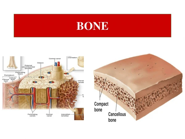
Physical Structure: Composed of cells and predominantly collagenous extracellular matrix (type I collagen) called osteoid which become mineralized giving bone rigidity and strength • Compact (cortical) bone • Dense rigid outer shell • Minimal gaps and spaces • Accounts for 80% of the total bone mass of an adult skeleton
Physical Structure: • Trabecular (cancellous) bone • Central zone of interconnecting trabeculae • Network of rod- and plate-like elements • Make the overall organ lighter • Allow room for blood vessels and marrow
Microscopic Structure: Woven: • Haphazard organization of collagen fibers • Mechanically weak • Produced when osteoblasts produce osteoid rapidly • E.g. Fetal bones, fractures, Paget’s
Microscopic Structure: Lamellar: • Regular parallel alignment of collagen into sheets • Mechanically strong • Fibers run in opposite directions in alternating layers • Replaces woven bone after fracture
Cellular Structure: • Derived from osteoprogenitor cells • The bone-forming cells • Synthesize osteoid, mediates its mineralization • Found lined up along bone surfaces Osteoblasts
Osteoblast Stimulation • Stimulated to increase bone mass through increased secretion of osteoid • Stimulated by the secretion of: • Growth Hormone • Thyroid Hormone • Sex Hormones (oestrogens + androgens) • These hormones also promote increased secretion of osteoprotegerin • Inhibits osteoclast stimulation
Osteoblast Stimulation • Vit D + PTH + Osteocytes stimulates osteoblasts to secrete cytokines: • Stimulate bone resorption via osteoclasts • Differentiation of progenitor cells to osteoclasts • Decrease Osteoprotegerin
Cellular Structure: • Derived from macrophage monocyte cell-line • Phagocytic cells • Responsible for bone resorption • Important along with osteoblasts in the constant turnover and refashioning of bone Osteoclasts
Osteoclast Inhibition • Rate of bone resorption inhibited by: • Calcitionin (C cells of thyroid) • Osteoprotegerin (osteoblasts)
Cellular Structure: • Mature bone cells • Inactive osteoblasts, trapped and surrounded by bone matrix • Function: • Formation of bone • Matrix maintenance • Calcium homeostasis Osteocytes
Bone matrix • Type I Collagen • Ground substance proteoglycans • Non-collagen molecules involved in mineralization regulation 70% inorganic salts, 30% organic matrix Organic matrix:
Bone matrix Type I collagen: Ground substance proteoglycans: • Polymer of numerous elongated overlapping tropocollagen subunits • Hole zones initial site of mineral deposition • Controls water content in bone • Regulating formation of collagen fibers in a form appropriate for mineralization
Bone matrix • Calcium and phosphate in form of hydroxyapatite Non-collagen molecules: Inorganic component: • Osteocalcin: • Binds calcium • Osteonectin: • bridging function between collagen and mineral component
How is bone formed? • Collagen synthesized by osteoblasts • Secreted as osteoid • After maturation phase • Amorphous calcium phosphate precipitates in hole zones • Mineralization foci expand + coalesce into hydroxyapatite crystals • 20% remains amorphous for readily available calcium buffer
How is bone formed? • Concentration of calcium + phosphate in extracellular fluid greater than required for spontaneous calcium deposition • Inhibited by pyrophosphate • Deposition of calcium controlled by osteoblasts which secretes alkaline phosphatase vesicles • Neutralizes pyrophosphate
Bone development and growth • Develops in 2 ways (2 types of ossification) • Both involve replacement of primitive collagenous supporting tissue by bone • Resultant woven bone is then extensively remodelled by resorption and appositional growth to form mature adult lamellar bone • Thereafter the process occurs at much reduced rate to accommodate functional stresses and to effect calcium homeostatis
Bone development and growth • Two types of occification: • Endochondral ossification • Intramembranous ossification
Endochondral ossification • E.g. long bones, vertebra, pelvis, base of skull • Hyaline cartilage is first formed in a shape corresponding closely to future bone • Cartilage model is covered - perichondrium • Bone matrix deposition - replacing the existing cartilage
Intramembranous ossification • E.g. vault of skull, maxilla, mandible • Deposition of bone in primitive mesenchymal tissue • Direct replacement of mesenchyme by bone • Cell differentiation into osteogenic tissue • These become impregnated with calcium salts
Remodeling/Bone turnover • Process of resorption followed by replacement of bone, with little change in shape • Occurs throughout a person's life • Purpose: • To regulate calcium homeostasis • Repair micro-damaged bones • To shape and sculpture the skeleton during growth
The role of bone in calcium homeostasis • Bone contains 99% of total body calcium • Bone resorption releases calcium into systemic circulation • Bone formation actively binds calcium, removing it from blood stream • Ca2+ homeostasis controlled by: • Parathyroid hormone (parathyroid glands) • Calcitonin (Thyroid) • Calcitriol (Vit D3)
The role of bone in calcium homeostasis • Increases serum Ca2+ • Increases bone resorption by osteoclasts indirectly • Mediated by paracrinese.g. osteoprotegerin • Enhances renal reabsorption of calcium • Increases intestinal absorption of calciam • Via effects on Vit D Parathyroid hormone:
The role of bone in calcium homeostasis • Released when plasma Ca2+ increases • Decreases bone resorption • Increases renal calcium excretion • Enhances intestinal absorption of calcium • Facilitates renal reabsorption • Helps mobilize Ca2+ out of bone Calcitonin Calcitriol
References: • Human Physiology an Integrated Approach • Dee UnglaubSilverthorn • Wheater’s Functional Histology • B. Young, J.W. Heath • en.wikipedia.org/wiki/Bone • July 2012