
JI bypass procedure
E N D
Presentation Transcript
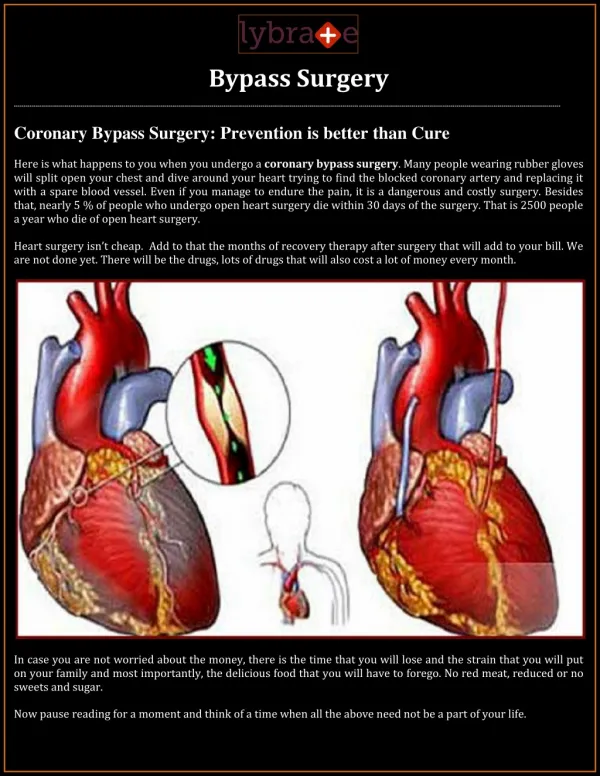
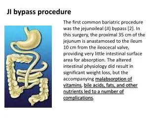
JI bypass procedure The first common bariatric procedure was the jejunoileal (JI) bypass [2]. In this surgery, the proximal 35 cm of the jejunum is anastamosed to the ileum 10 cm from the ileocecal valve, providing very little intestinal surface area for absorption. The altered intestinal physiology did result in significant weight loss, but the accompanying malabsorption of vitamins, bile acids, fats, and other nutrients led to a number of complications.
The tubular calcium oxalate deposits formed translucent crystals with an intraluminal, intracellular, and focally interstitial distribution (Figure 1, C and D)
Under polarized light, the deposits appeared strongly birefringent forming fan-like, sheaf-like, or irregular shapes. They were seen predominantly in the cortex and involved both proximal and distal tubules, with fewer deposits in the medulla
In general, intraluminal crystals predominated over intracellular and interstitial crystals. In many instances, the calcium oxalate crystals were associated with peritubular chronic inflammation, including four cases in which foreign-body giant cell reaction was seen
In all biopsies, the oxalate crystalline deposits were accompanied by diffuse acute tubular injury in non-atrophic tubules characterized by luminal ectasia, epithelial simplification, loss of proximal tubular brush border, enlarged reparative nuclei with prominent nuclei, and coarse cytoplasmic vacuolization (Figure 1D).
for patients with oxalate nephropathy after JI bypass, the mean time from surgery to renal failure was 44.5 mo (range 6.0 mo to 25 yr)
The prognosis of oxalate nephropathy after RYGB seems to be dismal, with progression to ESRD within 3 mo in 72.7% of patients in this study. Coexistent DGS or changes secondary to obesity and hypertension, present in 81.8% of patients, likely contributed to the poor outcomes.