Download
1 / 41
420 likes | 627 Views
Evaluation & Treatment of TMD. Presented by: Christy Dauner, OTR Laurie Applebee, PT Susan Vaughn, MS, OTR. Learning Objectives. Identify TMD risk factors and related diagnoses Differentiate joint and muscle disorders Understand goals of Occupational Therapy for TMD

E N D
Evaluation & Treatment of TMD Presented by: Christy Dauner, OTR Laurie Applebee, PT Susan Vaughn, MS, OTR
Learning Objectives • Identify TMD risk factors and related diagnoses • Differentiate joint and muscle disorders • Understand goals of Occupational Therapy for TMD • Understand OT treatments for TMD and muscle disorders • Perform assessment and treatment approaches for TMD
Disorders of the TMJ • Myofascial Dysfunction • Internal Derangement • Capsulitis • Subluxation • Arthritis
Risk Factors for TMD • Trauma such as blow to the jaw, whiplash injuries, MVA, dental work, opening the mouth too wide or for too long, prolonged chewing • Oral parafunctional habits such as clenching and bruxism that place continued strain on the masticatory system • Malocclusion causes bite instability or functional interference during chewing that places postural strain on the masticatory system • Stressful life events can trigger parafunctional habits and muscle guarding/tension • Emotional factors such as depression or anxiety decreases the ability to cope with pain and can increase parafunctional habits.
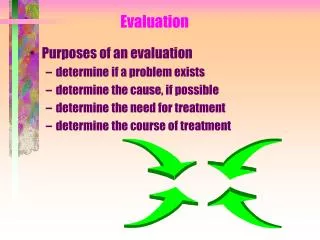
TMJ Evaluation • History & Symptoms (referred pain) • Functional Limitations • Tests, Measures & Palpation • AROM (active/passive incisal opening, lateral excursion, and protrusion) • PROM – scissor stretch • TMJ Noise • Muscle Palpation
Differential Diagnosis • Scissor stretch test: if opens further - muscular, if not - internal derangement • Clench test: bite down on tongue depressor for 10 – 15 seconds. Pain on same side – muscle, opposite side – joint • “S” vs. “C” curve with opening
Occupational Therapy Goals for TMD • Increase ROM to >40mm • Decrease pain • Teach joint protection (decrease parafunctional habits, limited opening) • Improve function (eating, yawning, DDS visit tolerance, oral hygiene, talking, sleep, work) • HEP independence • Neutral posture (head on neck, jaw, scapular position, TUTA)
Myofascial Pain Dysfunction • Most common disorder • Referred muscle pain • Muscle pain aggravated by jaw function or parafunction • HA’s • Tenderness of muscles w/o mechanical symptoms • Loss of motion or painful motion
Myofascial Pain Dysfunction • Caused by an underlying related disorder – malocclusion, arthritis, internal derangement, poor posture • Education is key! – posture, parafunction, stress management • Often chronic and cyclical • Often a myofascial component with all diagnoses
Myofascial Dysfunction Myofascial contributors may include: • * Lateral pterygoid * Medial pterygoid * Temporalis * Masseter * Digastrics * Muscles of the cervical spine
Lateral PterygoidOrigin: Lateral Pterygoid Plate of Sphenoid Insertion: Condylar Neck, Ramus of Mandible and Disc
TMJ Muscles – Lateral Pterygoid • #1 myofascial source of pain • Due to attachment to disc it can cause disc and jaw to be unable to return to normal resting position and cause clicking or popping. • Malocclusion of teeth/missing teeth
Medial PterygoidOrigin: Inner Surface of Lateral Pterygoid Plate Insertion: Ramus of Mandible by the Angle
TMJ Muscles – Medial Pterygoid • Stuffiness in ear • Swallowing difficulty as restriction in protrusion of jaw
Referral pattern – posterior mandible, mouth, below and behind TMJ including internal ear – not teeth
TemporalisOrigin: Temporal Fascia, Superior to Zygomatic ArchInsertion: Coronoid Process of Mandible
TMJ Muscles - Temporalis • Significant postural muscle (the only time it isn’t working is when you’re lying supine) • Perpetual clenching
Referral pattern – lower jaw, molar teeth and gum, maxilla, lower portion of mandible, temple eyebrow and external ear
Masseter Origin: Zygomatic ArchInsertion: Mandibular Angle and Ramus
TMJ Muscles - Masseter “Sinusitis”
Referral pattern - temple, along eyebrow, behind eye or upper teeth
DigastricsOrigin: Mastoid Notch (posterior), Symphysis of Mandible (anterior)Insertion: Join by a Common Tendon to the Hyoid Bone
TMJ Muscles - Digastrics • Rarely in spasm due to forward head posture (stretch weakness)
Referral pattern – behind mandible toward back of ear, lower incisors
Cervical Spine Muscles • Form stable base for TMJ on which to work • Poor posture – condyle rotates backward – change of biomechanics • Referral pattern from the cervical spine-Temporal Headaches, SCM • Assess for tension in upper traps, scalenes, and SCM
Parafunctional Behaviors • Gum/candy chewing (chewing limited to 15 – 20 minutes/day!) – including chewing on one side • Clenching/bruxing/grinding • Leaning on chin/jaw • Biting nails, pencils, cheeks • Sleep position • Caffeine use • Musical instruments • Mouth breathing • Phone cradling
Treatment – Myofascial Pain Dysfunction • Modalities: US - 1.0 – 1.2 w/cm2, 3 MHz, x5 minutes to joint or muscle, heat, electrical stimulation • Manual Therapy – joint mobs/distraction, MFR – including upper cervical region • HEP/Lifestyle changes • Tongue positioning (TUTA) • Self-joint distraction &/or MFR • Eliminating parafunctional behaviors • Postural instruction • Conjunction with splint therapy &/or counseling (Referral to psychology for CBT as needed for stress and anxiety management)
Disc DisorderInternal Derangement • Disc held in place by collateral ligaments and posterior ligament, w/ movement dictated by lateral pterygoid • Click, pop, lock • Pain at joint • “S” shaped opening/closing to reposition jaw • Eye pain • History of trauma
Treatment – Internal Derangement • Modalities: Iontophoresis, electrical stimulation, cold – ice massage • Manual therapy – Joint distraction • Joint protection techniques: Limit motion to no noise, soft food diet or chewing behaviors • Home exercise instruction • Change parafunctional behaviors • Self joint distraction techniques • Tongue positioning for relaxation (TUTA) • Postural instruction and controlled opening/neuromuscular re-education
Treatment - Other • Capsulitis • Usually a result of another disorder unless post surgery • Modalities, MT and HEP • Subluxation • Excess opening (>40 mm) • Usually a component of myofascial pain dysfunction, and treated as this, with addition of stab exercises and controlled opening
Treatment - Other • Arthritis • Generalized joint pain and inflammation • Usually seen in conjunction w/ other Dx • Joint protection, rest • Stretching, therapeutic exercise • Modalities (cold vs. heat, pulsed US, phono/iontophoresis, E-stim)
Intervention: Dentist • Assess occlusion • Parafunctions of clenching/bruxing • Malocclusions • Pressure on back teeth activate temporalis an superior head of lateral pterygoid, anterior teeth activatemasseters
Lab - Evaluation • AROM (Therabite) • Active Incisal Opening (Normal 40-60 mm) • Passive Incisal Opening (Normal 42-62 mm) • Lateral Excursion (Normal >7 mm) • Protrusion (Normal > 7 mm)
Lab - Evaluation • TMJ Palpation/Observation • Quality of Motion: Smooth/Rigid/Jerky/Guarded/Fasciculation/ Thrusting • TMJ Noise: Opening Click, Closing Click, Reproducible • Visually Assess Opening (S or C Shaped Curve)
Lab • Muscle Palpation • Medial Pterygoid (elevation, protrusion, and lateral deviation to opposite side) • Place index finer on muscle at inside of bottom teeth in mouth. Place opposite thumb under jaw line below ear. Apply pressure to muscle as if to touch finger and thumb. Move along gum line until reach incisors in front. Hold until relaxes 1-2X/day • Lateral Pterygoid (elevation, protrusion, and lateral deviation to opposite side) • Place index finger inside mouth, under cheek bone. Point finger up and towards opposite eye. Apply pressure to muscle until it relaxes. To check positioning of finger, actively move jaw in opposite direction and muscle will contract under finger. Hold until relaxes 1-2X/day
Lab • Manual Therapy • Trigger point release • Joint distraction • Place thumb on back, bottom molar and wrap fingers under jaw • Press down as you lift on jaw in scooping motion • Do NOT pull jaw forward
Thank You • Feel free to contact Christy at 952-908-2567 or at Christyd@pdrclinics.com with questions. • PDR Clinic Locations: Edina, Burnsville, Maplewood, Burnsville, Chanhassen • Specializing in the treatment of chronic neck, back and TMJ pain.