Distribution and Composition of Microplankton in the Southeastern Pacific Ocean
70 likes | 194 Views
This study explores the distribution and composition of microplankton across different oceanic regimes, from the eutrophic coastal upwelling of Chile to ultra-oligotrophic zones in the South Pacific gyre. It includes the investigation of temporal variations in distribution and the presence of nitrogen-fixing organisms such as Trichodesmium spp. Utilizing a combination of methodologies including light microscopy, fluorescence microscopy, scanning electron microscopy, and gene sequencing, this research aims to enhance understanding of marine phytoplankton taxonomy and biodiversity in under-researched oceanic areas.

Distribution and Composition of Microplankton in the Southeastern Pacific Ocean
E N D
Presentation Transcript
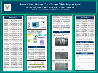
Title Distribution and composition of microplankton along the SE Pacific Ocean Fernando Gómez1* & Hervé Claustre2 1Department of Aquatic Biosciences,The University of Tokyo 2Laboratoire d’Océanographie deVillefranche-sur-Mer *address until February 2004 fernando.gomez@fitoplancton.com
To study the distribution (vertical and longitudinal) and composition of microplankton from the eutrophic (coastal upwelling of Chile) to ultra-oligotrophic regimes (South Pacific gyre). To study the temporal variations on the distribution (diel cycle) in selected stations Objectives To investigate the occurrence and distribution of N2-fixers (Trichodesmium spp.) and N2-fixer symbiotic associations (Richelia and others) To contribute to the study of marine phytoplankton taxonomy and biodiversity in one of the less studied major oceanic entities of the world ocean
Sample collection Niskin bottle 3 ml Lugol # station, depth 500 ml seawater Polyestyrene plastic bottle Storage: dark, cool and quiet place 0, 5, 10, 20, 30, 50, 70, 90, 100, 120, 150, 175, 200 m depth
Methodo Pre-concentration Methodology 500 ml Lugol fixed sea-water samples Settling in counting chambers Microscopical analysis (inverted microscope)
The specimens of interest will be isolated with a capillary from the chambers: Taxonomical studies: 1. Light microscopy High magnification microphotographs (×1000) Nomarski Differential Interference Contrast (D.I.C.) Morphology and location of organelles such as chloroplasts, pyrenoids, nuclei, cellulose thecal plates, flagella, etc transferred to a glass slide staining compounds (flourochromes: DAPI, Fluorescent Brightener, etc) Fluorescence microscopy (UV light or blue light according to the staining compound)
Scanning Electron Microscopy The specimens of interest will be isolated with a capillary from the chambers: Taxonomical studies: 2. SEM Takayama method Adhered on the poly-L-lysine coated glass plate rinse in distilled water; dehydrate through an ethanol series Ion spatter coating with Au or Au-Pd Scanning Electron Microscopy
Taxonomical studies: 3. Gene sequencing After to complete the morphological characterization of the species of interest, the forthcoming specimens will be picked and keep for single-cell molecular analysis (phylogeny) Discrepancies on the most appropriate fixative (methanol) for Polymerase Chain Reaction (PCR) appear in the literature The protocol of extraction is quite similar, the gene amplification protocol varies according to the instruments and reagents suppliers Nucleotide sequences of coding (small subunit (SSU), partial large subunit (LSU) (specific diversity) and internal transcribed spacer region (ITS1-5.8SSU-ITS2) parts of the rRNA operon (intraspecific diversity)