Download
1 / 10
100 likes | 288 Views
Phototoxic Maculopathy After Scleral Sutured IOL Implantation. NC CHO, DW LEE, EY KWEON, MJ KIM Department of Ophthalmology, Chonbuk National University Medical School, Korea. The authors do not have any commercial or proprietary interest in the subject matter of this paper. Purpose.

E N D
Phototoxic Maculopathy After Scleral Sutured IOL Implantation NC CHO, DW LEE, EY KWEON, MJ KIM Department of Ophthalmology, Chonbuk National University Medical School, Korea
The authors do not have any commercial or proprietary interest in the subject matter of this paper
Purpose • Macular phototoxicity is a rarely reported complication of ocular surgery including complicated cataract extraction, complicated anterior segment procedures, and vitrectomy. • An increased risk of phototoxicity is associated with an operating time greater than 100 minutes, focus, retinal temperature and drugs. • The aim of this study was to report and emphasize the features of phototoxic maculopathy occurring in patients who underwent scleral-sutured IOL implantation.
Method • The charts of 75 patients who underwent scleral-sutured intraocular lens implantation at Chonbuk National University Hospital between March 1995 and February 20078 were reviewed. • Twenty-one patients underwent combined 3 port pars plana vitrectomy and sutured IOL (vitrectomy group). • Fifty-four patients did not do vitrectomy (nonvitrectomy group). • All surgeries were performed using the same coaxial illuminated microscope.
Method • All diagnoses were confirmed by careful fundus examination and fluorescein angiography. • An angiographic archive was retrospectively surveyed and interpreted by a retinal specialist to identify eyes with lesions characterized as typical of retinal phototoxicity
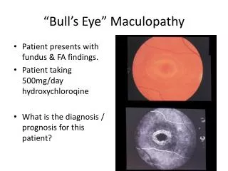
Results • Phototoxic maculopathies were detected in 7 of 75 sutured IOLs(9.33%). • Phototoxic maculopathy occurred more frequently in vitrectomy group(3eyes, 14.29%) than nonvitrectoy group(4eyes, 7.41%). • Immediately postoperatively, the patients reported decreased vision, purple to red color, scotoma • In 5 of the phototoxic maculopathy cases(71.43%), the visual acuity was 20/200 or worse (table 1). • Fluorescein angiography showed coarse alterations of the retinal pigment epithelium(figure1,2).
Results • Fundus photographs show irregular, non elevated oval shaped retinal pigmented lesion and fluorescein angiograms show a irregular hyperfluorescence involving macula with no vascular leakage nor edema. (Figure 1)
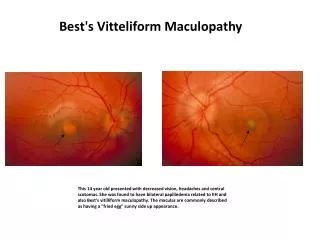
Results • Fundus photography show well-demarcated green-colored, crescent shape degeneration involving macula. Fluorescein angiograms show marginal hyperfluorecence of RPE atrophy with central hypofluoroscence for choroidal ischemic infarction. (Figure 2)
Conclusion • Phototoxic maculopathy is a rarely reported complication of intraocular surgery including scleral fixation, vitrectomy • Foveal injuries after this surgery are more profound with poor visual outcome. • The most important factor in performing surgery is to keep the focused illumination beam off the central fovea and limit exposure times as much as practical. • Much work still needs to be done to assess the risk of iatrogenic photoretinal injuries and methods of prevention. The theoretical advantages of filters, indirect illumination system,and corneal shield need to be prospectively evaluated againtst a control group to determine their efficacy.