Airway
Chapter 7. Airway. Case History.

Airway
E N D
Presentation Transcript
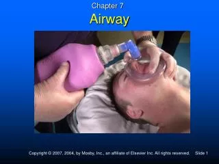
Chapter 7 Airway
Case History You respond to a motor vehicle crash. On arrival, you find a demolished vehicle with a 40-year-old unresponsive, cyanotic male slumped over the wheel of his car. He is making gurgling noises as he breathes shallowly at a rate of 8 times per minute. Blood and loose teeth are in the upper airway.
Analysis of the Case • Rapid extrication with immobilization of cervical spine • Airway: jaw thrust, suction, oropharyngeal airway • Mouth-to-mask or bag-valve-mask with supplemental oxygen, or flow-restricted, oxygen-powered ventilator • Continuously monitor signs of adequate ventilation and oxygenation
Overview • Management of the airway and ventilation represent the first and most critical steps in most serious emergencies. • Humans are oxygen-dependent organisms. • Three critical priorities • Airway • Breathing • Oxygenation
Assess the Airway, Breathing, Circulation in Every Patient • Assess for adequate vs. inadequate breathing to determine if the patient needs treatment to: • Maintain airway • Support ventilation • Supplement oxygenation
Airway Interventions • Manual techniques • Head tilt/chin lift • Jaw thrust • Mechanical techniques • Oropharyngeal airways • Nasopharyngeal airways • Suction
Ventilation Interventions • Mouth-to-mask • Bag-valve-mask • Flow-restricted oxygen-powered ventilator
Oxygen Interventions • Supplemental oxygen for patient with adequate ventilation • Nonrebreather • Nasal cannula (when patient cannot tolerate mask) • Blow-by oxygen for children (when patient cannot tolerate mask) • Positive pressure • Mouth-to-mask with supplemental oxygen • Bag-valve-mask with supplemental oxygen • Flow-restricted oxygen-powered ventilator
Review of Anatomy and Physiology • Three main functions of respiratory system • Delivery of oxygen from atmosphere to blood • Removal of carbon dioxide from the blood to the atmosphere • Creation of voice
Structures and Function • Nose • Pharynx • Epiglottis and larynx • Trachea • Bronchi and bronchioles • Alveoli
Lungs • Two cone-shaped organs consisting of: • Bronchi, bronchioles, and alveoli • Suspended in thoracic cavity and separated by mediastinum • Surrounded and protected by ribs thoracic spine, sternum, clavicle, and muscles • Outer surface of lungs and inner surface of thoracic cavity lined with pleura
Elasticity of the Lungs • Lungs have natural tendency to collapse – “elasticity.” • Lungs fill thoracic cavity because of relative “negative” pressure between pleura. • Pneumothorax disturbs relationship; lung collapses
Muscles of Breathing • Cause chest movement that results in air exchange • Quiet breathing involves two muscles: • Diaphragm • External intercostals
Inspiration (Active Process) • Diaphragm increases inferior-superior diameter of chest cavity • External intercostals increase anterior-posterior, lateral dimensions • Increase in size results in more volume, less pressure relative to atmosphere, and air rushes in. • Inspiration continues until pressure within lung and atmosphere equalizes.
Expiration (Passive Process) • Respiratory muscles relax and elastic recoil of lungs. • Chest cavity decreases in size - less volume, pressure increases relative to atmosphere. • Air exits airway until atmosphere and chest pressure are equal.
Accessory Muscles of Breathing • Needed for forceful breathing • Key sign of respiratory distress • Accessory muscles of inspiration increase the size of chest cavity by further lifting rib cage and increasing diameter • Inspiratory muscles – scalene (neck), sternocleidomastoids, parasternal (chest)
Accessory Muscles of Expiration • Assist in forcefully evacuating air • Pull down on ribs and compress abdominal contents into diaphragm • Expiratory muscles — internal intercostals and abdominal muscles • Active accessories indicate respiratory distress, signs include bulging neck muscles, retraction between ribs, abdominal distention.
Respiratory Volumes • Minute volume • The amount of air delivered to the lungs each minute • Equals tidal volume times the number of breaths per minute • Normal minute volume for an adult • Tidal volume (approx 500 mL/breath) x respiratory rate (normal range is 12-20/minute) • Example: MV = 500 mL/breath x 12 breaths/min or 6000 mL/min
Using Minute Ventilation An unresponsive male has respirations of 6 per minute and no noticeable chest rise. Is his minute ventilation adequate to support life?
Using Minute Ventilation • Provide rescue breathing to the patient with hypoventilation. • How do you gauge adequate volume per breath? What is the tidal volume? • How many breaths a minute should be administered? • What is the minute ventilation?
Ventilation • Adequate ventilation • Respiratory rate of 12 - 20 with visible chest rise • Alert mental state, muscle tone, and moving air • Normal skin color • Inadequate ventilation (respiratory failure) • Very slow ventilation • Very rapid ventilation with minimal or no chest rise • Altered mental state, poor muscle tone, poor air flow, cyanosis
Alveolar-Capillary Exchange • Diffusion • Movement of gases from an area of higher concentration to an area of lower concentration • Oxygen and carbon dioxide diffuse at the lungs and at the tissues. • Lungs: oxygen moves from the alveoli (higher) to the blood (lower) • Tissues: oxygen moves from the blood (higher) to the tissues (lower) • Fluid in the alveoli can impair diffusion and lead to hypoxia.
Clinical Application Nerve agents, such as sarin, cause muscle paralysis and excessive secretions, including in the lungs. Victims die a respiratory death, from hypoventilation and/or “drowning in their own secretions.”
Neuroregulation of Breathing • CNS monitors • Carbon dioxide, oxygen, and pH • Directs respiratory muscles to rate and depth of ventilation, as needed • Carbon dioxide primary drive for normal person • Patients with COPD • Oxygen may be primary drive for COPD • Use caution when administering oxygen • Be prepared to provide positive-pressure ventilation if a COPD patient begins to hypoventilate.
Infant and Child Considerations • Mouth and nose —smaller • Obstructed more easily • More space taken up by tongue • Lower tidal volumes • More prone to gastric inflation
Adequate Breathing • Rhythm • Regular • Irregular • Quality • Breath sounds — present and equal • Chest expansion — adequate and equal • Minimum effort of breathing • Depth (tidal volume) – visible chest rise
Adequate Breathing Rate • Adult — 12-20/minute • Child — 15-30/minute • Infant — 25-50/minute
Important Terms • Respiratory distress • Respiratory failure • Respiratory arrest
Respiratory Distress • Shortness of breath • Agitation or restlessness • Active accessory muscle use • Retractions • Cyanotic skin • Increased pulse rate • Increased respiratory rate
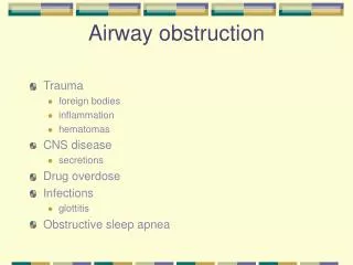
Noisy Breathing • Crowing • Audible wheezing • Gurgling • Snoring • Stridor • A harsh sound heard during breathing • Upper airway obstruction
Respiratory FailureInadequate Breathing • Depressed mental state (e.g., responsive to voice, responsive to pain, or unresponsive) • Rate – very fast or very slow • Rhythm — irregular • Quality • Breath sounds — diminished or absent • Chest expansion — unequal or inadequate • Depth (tidal volume) — inadequate/shallow • Cyanotic skin color
Inadequate Breathing • Rate — outside of normal ranges • Rhythm — irregular • Quality • Breath sounds — diminished or absent • Chest expansion — unequal or inadequate • Increased effort of breathing — use of accessory muscles — predominantly in infants and children • Depth (tidal volume) — inadequate/shallow
Respiratory Arrest Definition: Complete cessation of breathing
Airway Interventions • Manual techniques • Head tilt/chin lift • Jaw thrust • Mechanical techniques • Oropharyngeal airways • Nasopharyngeal airways • Suctioning
Opening the Airway • Head tilt/chin lift • Jaw thrust (if injury to the neck is suspected)
Techniques of Suctioning • Body substance isolation • Purpose • Remove blood, liquids, and food particles from the airway • Some units are inadequate for removing solid objects • Suction immediately when a gurgling sound is heard
Soft (French) • Useful for suctioning the nasopharynx • When rigid device is not possible • Inserted only as far as the base of the tongue
Suctioning Issues • Suction for no more than 15 seconds at a time (less in infants and children). • Secretions that cannot be removed quickly • Log roll, clear manually • If patient produces frothy secretions as rapidly as suctioning can remove: • Suction for 15 seconds • Artificially ventilate for 2 minutes • Suction for 15 seconds • Continue in that manner • Rinse catheter and tubing as needed.
Oropharyngeal Airway • Measure the airway.
Oropharyngeal Airway • Insert airway upside down, with the tip facing the roof of the patient’s mouth. • Rotate device into position. • Alternate method: Hold the tongue down and forward with a tongue depressor and insert airway directly.
Nasopharyngeal Airway • Measure from tip of nose to tip of ear. Look at diameter of nostril.
Nasopharyngeal Airway Insert airway with bevel toward base of nose or septum.
Positive-Pressure Ventilation • Required for patients in respiratory failure and respiratory arrest • Characteristics of effective positive-pressure ventilation • Rate appropriate for age • Adult – One breath every 5-6 seconds • Children and infants – One breath every 3-5 seconds • Chest rise observed with each breath
Positive-Pressure Ventilation • Characteristics of effective positive-pressure ventilation (continued) • Each breath delivered slowly • Over 1 to 2 seconds for adults • Over 1 to 1.5 second for children and infants • Oxygen saturation is greater than 93%, if pulse oximeter is used.
Positive-Pressure VentilationOrder of Effectiveness • Mouth-to-mask • Two-person bag-valve-mask • Flow-restricted, oxygen-powered ventilation device • One-person bag-valve-mask
Mouth-to-Mask • Allows for good seal and for direct feedback to the rescuer
Bag-Valve-Mask • Oxygen reservoir • Self-refilling bag • Valve to allow oxygen inlet flow of 15 L/min • Valve to make it nonrebreather • If pop-off valve, ability to disable if necessary when high resistance encountered
Bag-Valve-Mask • Bag capacity — approximately 1,600 mL • Single rescuer may have difficulty maintaining an airtight seal. • Result — May provide less volume than mouth-to-mask
Bag-Valve-Mask • Select correct mask size (adult, infant or child). • Position thumbs over top half of mask. • Place index and middle fingers over bottom half. • Place apex of mask over bridge of nose. • Lower mask over mouth and upper chin. • If mask has large round cuff, position center port over mouth.