Download

1 / 20
210 likes | 258 Views
Learn about the parasite Dirofilaria immitis causing heartworm disease in dogs and cats, its life cycle, clinical signs, diagnosis, treatment, prevention, and the incidence in different species.

E N D
Heartworm disease in the dog was first recognized and described in a veterinary journal published in 1847. • Feline heartworm disease was described in 1922.
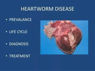
The Parasite • HW Dz is caused by the nematode Dirofilaria immitis • This is a large worm that lives throughout the body during various life stages, but the adult worms are housed in the right side of the heart and in the pulmonary artery.
Adults of both sexes live in the heart, where they mate and the female produces microfilariae • Female heartworms can be 14” long • Microfilariae are released into the blood stream
Quick Lifecycle Review • Adults produce microfilariae and release them into bloodstream. (L1) • Mosquito suck up blood and microfilariae during a blood meal (over next 2 weeks, microfilariae molt into L2, then L3) • Mosquito bites dog and now releases infective L3s into tissues of bitten animal • L3s migrate through tissues, molting twice. (L4 & L5) • L5s reach heart, gain sexual maturity, and reproduce
Clinical signs • Most signs reflect heart or lung damage • Cough • Exercise intolerance • Abnormal lung sounds • Heart enlargement • Ascites
Severity of disease depends on: • Length of infection • Number of worms present • Size of affected animal • Activity level of affected animal
Some affected species • All canids • Domestic and wild cats • Ferrets and other mustelidae • Sea lions • Humans
Human infection • Human heartworm infections can occur, but they are never patent • Microfilariae encyst in tissues • Typically, “coin lesions” are found during an x-ray exam • Most cases are asymptomatic • Nodules in the chest are surgically removed since they could be cancerous masses
Diagnosis in Dogs • Changes to the heart and/or lungs might be seen on • Radiographs • Ultrasound • Echocardiography
Microscopic exams • Direct smear: may or may not detect microfilaria in a positive sample • Results are only meaningful if positive! • Knott’s test: more accurate but false negatives can occur • single sex infections • immune system interference • You must differentiate D. immitis from Dipetalonema reconditum
Serology • Previous serological tests were antibody tests • Most recent tests are antigen tests • Detect adult heartworm antigen • Reveals occult infections (when no microfilaria are present)
Treatment • Two phases: • Adulticide • Larvacide • Adulticide: • Old: thiacetarsemide • New: melarsomine dihydrochloride • Larvacides: • Macrocyclic lactones
Post-treatment testing • Antigens will be gone in about 4 months
Prevention • Diethyl carbamizine (Filaribits) • Sentinal (milbemycin oxime) • Revolution (sealamectin) • Proheart (moxidection) • Heartgard, Iverhart (ivermectin)