Download
1 / 39
390 likes | 511 Views
Understand types, clinical features, and management of meningitis in a 30-year-old male with fever, vomiting, headache, and unconsciousness. Learn about subacute sclerosing panencephalitis and brain abscess basics. Presented by Dr. Ravi Vaswani.

E N D
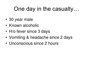
One day in the casualty… • 30 year male • Known alcoholic • H/o fever since 3 days • Vomiting & headache since 2 days • Unconscious since 2 hours
Acute CNS Infections Dr. Ravi Vaswani MD Professor, Department of Medicine Yenepoya Medical College, Mangalore
Objectives • By the end of this session, you will be able to know the • Common types and etiologies for meningitis • Clinical features of meningitis • Management of meningitis • Basics of subacute sclerosing panencephalitis • Basics of brain abscess
Background • Early recognition, efficient decision-making, and rapid institution of therapy can be lifesaving • Distinct clinical syndromes • acute bacterial meningitis • viral meningitis, encephalitis • focal infections: brain abscess & subdural empyema • infectious thrombophlebitis • Chronic meningitis • Thought to be benign, until altered consciousness, focal neurologic signs, or seizures appear
Temporal profile classification • Acute (pyogenic) • Hours to days • Subacute/Chronic (basal) • More than 14 days
Bacterial meningitis • Bacterial meningitis is an acute purulent infection within the subarachnoid space • CNS inflammatory reaction results in decreased consciousness, seizures, raised intracranial pressure (ICP), and stroke • The meninges, the subarachnoid space, and the brain parenchyma are all frequently involved in the inflammatory reaction (meningoencephalitis).
Epidemiology • Organisms most commonly responsible for community-acquired bacterial meningitis • Streptococcus pneumoniae (~50%) • N. meningitidis (~25%) • group B streptococci (~15%), and • Listeria monocytogenes (~10%) • H. influenzae now accounts for <10% of cases of bacterial meningitis
S. pneumoniae • Most common cause of meningitis in adults >20 years of age • Risk factors include • coexisting acute or chronic pneumococcal sinusitis or otitis media or pneumonia • alcoholism, diabetes • splenectomy, hypogammaglobulinemia, complement deficiency, and • head trauma with basilar skull fracture and CSF rhinorrhea
N. meningitidis • Accounts for 25% of bacterial meningitis • Up to 60% of cases in children and young adults between the ages of 2 and 20 • Petechial or purpuric skin lesions can provide an important clue
Others • S aureus and coagulase-negative staphylococci occurs following invasive neurosurgical procedures, particularly shunting procedures for hydrocephalus • L. monocytogenes has become an increasingly important cause of meningitis in neonates (<1 month of age), pregnant women, individuals >60 years, and immunocompromised individuals
Pathophysiology • Colonize nasopharynx by attaching to nasopharyngeal epithelial cells • Invade intravascular space by creating separations in apical tight junctions of columnar epithelial cells • Blood stream invasion • Infect choroid plexus epithelial cells, and gain access to the CSF
Pathophysiology • Many neurologic manifestations & complications result from immune response to pathogen rather than from bacteria-induced tissue injury (elevated levels of CSF cytokines and chemokines - TNF α, IL-1, 6, 12) • Neurologic injury can progress even after CSF is sterilized by antibiotic therapy • Inflammation rapidly increases when lysis of bacteria occurs
Clinicopathological correlation • Fever due to cytokines (TNF, IL) • Vomiting CTZ stimulation • Headache cerebritis & vascular changes • Vasculitis results in ischemia and infarction, obstruction of branches of the middle cerebral artery by thrombosis, thrombosis of the major cerebral venous sinuses, and thrombophlebitis of the cerebral cortical veins • Combination of interstitial, vasogenic, and cytotoxic edema leads to raised ICP and coma
Clinical features • Classic clinical triad of meningitis is fever, headache, and nuchal rigidity • Decreased level of consciousness occurs in >75% • Nausea, vomiting, and photophobia - common • Seizures - initial presentation or during the course of illness
Raised ICP • Headache, vomiting, blurring of vision, ataxia • Deteriorating or reduced level of consciousness • Papilledema • Dilated poorly reactive pupils • Sixth nerve palsy • Decerebrate posturing • Cushing reflex (bradycardia, hypertension, and irregular respirations) • Cerebral herniation
Investigations • Blood cultures • Fundus examination • CT, MRI • CSF examination via Lumbar puncture • Guarded LP
Treatment • A medical emergency • Goal is to begin antibiotic therapy within 60 min of a patient's arrival in the emergency room • Empirical antimicrobial therapy • S. pneumoniae and N. meningitidis are the most common etiologic organisms of community-acquired bacterial meningitis • Due to emergence of penicillin- and cephalosporin-resistant a combination of dexamethasone, a third-generation cephalosporin (e.g., ceftriaxone or cefotaxime) and vancomycin
In resource limited setting… • Inj Crystalline Penicillin 20 lakh units every 2 hrs • Inj Gentamicin 60-80 mg q8h • Inj Metronidazole 500 mg q8h
Adjunctive therapy • Glucocorticoids: Inj Dexamethasone 10 mg IV 20 min before or with the first dose of antibiotic • Not later than 6 hr after first antibiotic dose • Treatment of raised ICP: elevation of the patient's head to 30–45°, intubation and hyperventilation (PaCO2 25–30 mmHg), and mannitol/furosemide
Chronic Meningitis • Symptoms more than 14 days • Less prominent symptoms • Fever often absent
Etiology • Partially treated bacterial meningitis • M. tuberculosis • Fungal: C. neoformans, H. capsulatum, C. immitis • T. pallidum • Viral (HIV, etc) • Aseptic meningitis • Mollaret’s meningitis
CSF abnormalities • Elevated opening pressure • Lymphocytic pleocytosis (10–500 cells/ L) • Elevated protein concentration in the range of 1–5 g/L (10–500 mg/dL) • Decreased glucose concentration in the range of 1.1–2.2 mmol/L (20–40 mg/dL) • Cobweb suggestive of TB
Treatment • AntiTB • Amphotericin for cryptococcal or other fungal + lifelong fluconazole • Acyclovir if viral suspected • Shotgun therapy; Blunderbuss therapy
Subacute Sclerosing Panencephalitis (SSPE) • Rare chronic, progressive demyelinating disease of associated with a chronic nonpermissive infection of brain tissue with measles virus • Estimated at 1 in 100,000–500,000 measles cases • Average of 5 cases/year reported in United States • Most give h/o primary measles infection at early age (2 yrs) • Latent interval 6–8 years; • 85% between 5 and 15 years old at diagnosis • Initially poor school performance & mood & personality changes • Typical signs of fever & headache, absent • As disease progresses, pts develop progressive intellectual deterioration, myoclonus, ataxia, and visual disturbances • Last, pts unresponsive, quadriparetic, & spastic, with hyperactive tendon reflexes & extensor plantar responses.
SSPE (Contd) • MRI is often normal • EEG periodic pattern with bursts of high-voltage, sharp, slow waves every 3–8 s, followed by periods of attenuated ("flat") background • CSF is acellular with mildly elevated protein concentration and a markedly elevated gamma globulin level (>20% of total CSF protein) • CSF antimeasles antibody levels are elevated. Measles virus can be cultured from brain tissue using special cocultivation techniques. • No definitive therapy for SSPE is available • Treatment with isoprinosine (100 mg/kg per day), alone or in combination with intrathecal or intraventricular α interferon, doubtful
Brain Abscess • A brain abscess is a focal, suppurative infection within the brain parenchyma, typically surrounded by a vascularized capsule. The term cerebritis is often employed to describe a nonencapsulated brain abscess • Relatively uncommon intracranial infection
Etiology • May develop by • contiguous spread from a cranial site of infection, such as paranasal sinusitis, otitis media, mastoiditis, or dental infection • following head trauma or a neurosurgical procedure • hematogenous spread from a remote site of infection • In up to 25% of cases, no obvious primary source of infection is apparent (cryptogenic brain abscess)
Pathogenesis • For bacterial invasion of brain parenchyma to occur, there must be preexisting or concomitant areas of ischemia, necrosis, or hypoxia in brain tissue • The intact brain parenchyma is relatively resistant to infection
Early cerebritis stage (days 1–3): characterized by perivascular infiltration, central coagulative necrosis • Late cerebritis stage (days 4–9): pus formation, enlargement of necrotic center, surround inflammatory infiltrate of macrophages and fibroblasts. A thin capsule of fibroblasts and reticular fibers develops, & surrounding area of cerebral edema becomes more distinct • Early capsule formation stage (days 10–13): better developed capsule on the cortical side. Correlates with appearance of ring-enhancing capsule on CT • Late capsule formation stage (day 14 and beyond): dense collagenous capsule. Cerebral edema regressed, marked gliosis outside the capsule
Clinical • Classic clinical triad of headache, fever, and a focal neurologic deficit • Hemiparesis is the most common localizing sign of a frontal lobe abscess • Temporal lobe abscess may present with a disturbance of language (dysphasia) or an upper homonymous quadrantanopia • Nystagmus and ataxia are signs of a cerebellar abscess • Signs of raised ICP
Treatment • CT/MRI for diagnosis • High-dose parenteral antibiotics and neurosurgical drainage • Third-generation cephalosporin (e.g., cefotaxime or ceftriaxone) and metronidazole • Aspiration and drainage of the abscess under stereotactic guidance • Prophylactic anticonvulsant therapy for 3 months after resolution