Fungi Chapter 31
950 likes | 997 Views
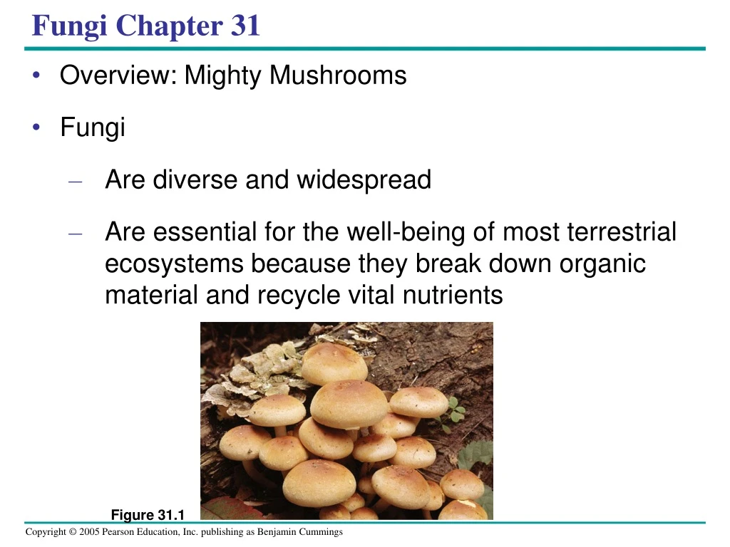
Figure 31.1. Fungi Chapter 31. Overview: Mighty Mushrooms Fungi Are diverse and widespread Are essential for the well-being of most terrestrial ecosystems because they break down organic material and recycle vital nutrients. Decomposers.

Fungi Chapter 31
E N D
Presentation Transcript
Figure 31.1 Fungi Chapter 31 • Overview: Mighty Mushrooms • Fungi • Are diverse and widespread • Are essential for the well-being of most terrestrial ecosystems because they break down organic material and recycle vital nutrients
Decomposers • Fungi are well adapted as decomposers of organic material • Performing essential recycling of chemical elements between the living and nonliving world
Nutrition Despite their diversity • Fungi share some key traits • Fungi are heterotrophs • But do not ingest their food • Fungi secrete into their surroundings exoenzymes that break down complex molecules • And then absorb the remaining smaller compounds
Fungi exhibit diverse lifestyles • Decomposers • Parasites • Mutualistic symbionts
Fungi consist of • Mycelia, networks of branched hyphae adapted for absorption • Most fungi • Have cell walls made of chitin
Reproductive structure.The mushroom produces tiny cells called spores. Hyphae. The mushroom and its subterranean mycelium are a continuous network of hyphae. Spore-producing structures 20 m Mycelium Figure 31.2 Body Structure • The morphology of multicellular fungi • Enhances their ability to absorb nutrients from their surroundings
Some fungi • Have hyphae divided into cells by septa, with pores allowing cell-to-cell movement of materials • Coenocytic fungi • Lack septa Cell wall Cell wall Nuclei Pore Septum Nuclei (a) Septate hypha (b) Coenocytic hypha Figure 31.3a, b
Nematode Hyphae 25 m (a) Hyphae adapted for trapping and killing prey Plant cell wall Fungal hypha Plant cell Plant cell plasma membrane (b) Haustoria Haustorium Figure 31.4a, b • Some unique fungi • Have specialized hyphae that allow them to penetrate the tissues of their host
Fungi produce spores through sexual or asexual life cycles • Fungi propagate themselves • By producing vast numbers of spores, either sexually or asexually
Key Heterokaryotic stage Haploid (n) Heterokaryotic (unfused nuclei from different parents) PLASMOGAMY (fusion of cytoplasm) Diploid (2n) KARYOGAMY (fusion of nuclei) Spore-producing structures Zygote SEXUAL REPRODUCTION Spores ASEXUAL REPRODUCTION Mycelium MEIOSIS GERMINATION GERMINATION Spore-producing structures Spores Figure 31.5 • The generalized life cycle of fungi
Sexual Reproduction • The sexual life cycle involves • Cell fusion, plasmogamy • Nuclear fusion, karyogamy • An intervening heterokaryotic stage • Occurs between plasmogamy and karyogamy in which cells have haploid nuclei from two parents • The diploid phase following karyogamy • Is short-lived and undergoes meiosis, producing haploid spores
2.5 m Figure 31.6 • Many fungi that can reproduce asexually • Grow as mold, sometimes on fruit, bread, and other foods
10 m Parent cell Bud Figure 31.7 • Other asexual fungi are yeasts • That inhabit moist environments • Which produce by simple cell division
Many molds and yeasts have no known sexual stage • Mycologists have traditionally called these deuteromycetes, or imperfect fungi
Fungi descended from an aquatic, single-celled, flagellated protist • Systematists now recognize Fungi and Animalia as sister kingdoms • Because fungi and animals are more closely related to each other than they are to plants or other eukaryotes
The Origin of Fungi • Molecular evidence • Supports the hypothesis that fungi and animals diverged from a common ancestor that was unicellular and bore flagella • Fungi probably evolved • Before the colonization of land by multicellular organisms
50 m • The oldest undisputed fossils of fungi • Are only about 460 million years old Figure 31.8
Arbuscular mycorrhizal fungi Zygote fungi Club fungi Sac fungi Chytrids Zygomycota Ascomycota Basidiomycota Glomeromycota Chytridiomycota Figure 31.9 • The phylogeny of fungi
Table 31.1 • A review of fungal phyla
Symbionts • Fungi form symbiotic relationships with • Plants, algae, and animals • Mycorrhizae • Are mutually beneficial relationships between fungi and plant roots • 90% of all plant species have mycorrhizae relationships with fungi.
The Move to Land • Fungi were among the earliest colonizers of land • Probably as symbionts with early land plants • The mycorrhizal symbiosis often increases the plant's uptake of inorganic compounds, such as nitrate and phosphate from soils • The fungal partners may also mediate plant-to-plant transfer of carbohydrates and other nutrients.
EXPERIMENT Researchers grew soybean plants in soil treated with fungicide (poison that kills fungi) to prevent the formation of mycorrhizae in the experimental group. A control group was exposed to fungi that formed mycorrhizae in the soybean plants’ roots. The soybean plant on the left is typical of the experimental group. Its stunted growth is probably due to a phosphorus deficiency. The taller, healthier plant on the right is typical of the control group and has mycorrhizae. CONCLUSION These results indicate that the presence of mycorrhizae benefits a soybean plant and support the hypothesis that mycorrhizae enhance the plant’s ability to take up phosphate and other needed minerals. Figure 31.21 Mycorrhizae • Mycorrhizae • Are enormously important in natural ecosystems and agriculture • Increase plant productivity RESULTS RESULTS
Fungus-Animal Symbiosis • Some fungi share their digestive services with animals • Helping break down plant material in the guts of cows and other grazing mammals
Figure 31.22 • Many species of ants and termites • Take advantage of the digestive power of fungi by raising them in “farms”
(a) A fruticose (shrub-like) lichen Figure 31.23a–c (b) A foliose (leaf-like) lichen (c) Crustose (crust-like) lichens Lichens • Lichens • Are a symbiotic association of millions of photosynthetic microorganisms held in a mass of fungal hyphae
Ascocarp of fungus Soredia Fungal hyphae Algal layer Algal cell Fungal hyphae Figure 31.24 10 m • The fungal component of a lichen • Is most often an ascomycete • Algae or cyanobacteria • Occupy an inner layer below the lichen surface
(b) Tar spot fungus on maple leaves (a) Corn smut on corn (c) Ergots on rye Figure 31.25a–c Pathogens • About 30% of known fungal species • Are parasites, mostly on or in plants
Practical Uses of Fungi • Some of the fungi that attack food crops • Are toxic to humans • Genetic research on fungi • Is leading to applications in biotechnology • Humans eat many fungi • And use others to make cheeses, alcoholic beverages, and bread
Staphylococcus Penicillium Zone of inhibited growth Figure 31.26 • Antibiotics produced by fungi • Treat bacterial infections
Figure 33.1 Chapter 33 Invertebrates • Overview: Life Without a Backbone • Invertebrates • Are animals that lack a backbone • Account for 95% of known animal species
How Animals are Built • Nearly all animals follow a physical plan. There are three kinds.
Asymmetry • Asymmetrical organisms do not develop complex communication, sensory or motor functions • The arrangement of body parts without central axis or point.
Radial Symmetry • The arrangement of body parts such that any plane passing through the oral-aboral axis divides the animal into mirror images • Allows for better specialization of sensory feeding and motor structure
Bilateral Symmetry • The arrangement of body parts such that a single plane passing through the oral-aboral axis divides the animal into mirror images • Allows for advanced specialization of sensory, feeding and motor function which usually occurs in a distinct head
Porifera Cnidaria Chordata Echinodermata Other bilaterians (including Nematoda, Arthropoda, Mollusca, and Annelida) Deuterostomia Bilateria Eumetazoa Ancestral colonial choanoflagellate Figure 33.2 • A review of animal phylogeny
Phylum Porifera • Sponges are sessile and have a porous body and choanocytes • Cellular – lack tissuesponges have no germ layers, no symmetry, no cephalization, no body cavity, and no segmentation • Reproduce sexually or asexually5000 known species • Sponges, phylum Porifera • Live in both fresh and marine waters
Choanocytes. The spongocoel is lined with feeding cells called choanocytes. By beating flagella, the choanocytes create a current that draws water in through the porocytes. 5 Flagellum Food particles in mucus Choanocyte Collar Azure vase sponge (Callyspongia plicifera) Osculum Spongocoel. Water passing through porocytes enters a cavity called the spongocoel. 4 Phagocytosis of food particles Amoebocyte Porocytes. Water enters the epidermis through channels formed by porocytes, doughnut-shaped cells that span the body wall. 3 The movement of the choanocyte flagella also draws water through its collar of fingerlike projections. Food particles are trapped in the mucus coating the projections, engulfed by phagocytosis, and either digested or transferred to amoebocytes. 6 Spicules Epidermis. The outer layer consists of tightly packed epidermal cells. 2 Water flow Amoebocyte. Amoebocytes transport nutrients to other cells of the sponge body and also produce materials for skeletal fibers (spicules). 7 Mesohyl. The wall of this simple sponge consists of two layers of cells separated by a gelatinous matrix, the mesohyl (“middle matter”). 1 • Sponges are suspension feeders • Capturing food particles suspended in the water that passes through their body Figure 33.4
Phylum Cnidaria • Jellyfish, sea anemones, coral • Radial symmetry - polyps & medusa • Bodies contain “jelly” (mesoglea) • Specialized stinging cells Sea Anemone
Tissue - lack organshave radial symmetry and 2 germ layers • lack cephalization, body cavity, & segmentation • Reproduce sexually or asexually11,000 known species4 groups - hydroids, box jellyfish, jellyfish, corals/anemones
The basic body plan of a cnidarian • Is a sac with a central digestive compartment, the gastrovascular cavity • A single opening • Functions as both mouth and anus
Medusa Mouth/anus Polyp Tentacle Gastrovascular cavity Gastrodermis Mesoglea Epidermis Body stalk Tentacle Mouth/anus Figure 33.5 • There are two variations on this body plan • The sessile polyp and the floating medusa
Prey Tentacle “Trigger” Discharge Of thread Nematocyst Coiled thread Cnidocyte Figure 33.6 • Cnidarians are carnivores • That use tentacles to capture prey • The tentacles are armed with cnidocytes • Unique cells that function in defense and the capture of prey
(d) Sea anemones and othermembers of class Anthozoaexist only as polyps. (b) Many species of jellies (classScyphozoa), including thespecies pictured here, are bioluminescent. The largest scyphozoans have tentaclesmore than 100 m long dangling from a bell-shaped body up to 2 m in diameter. (c) The sea wasp (Chironex fleckeri) is a member of class Cubozoa. Its poison,which can subdue fish andother large prey, is more potent than cobra venom. (a) These colonial polyps are members of class Hydrozoa. Figure 33.7a–d • Hydrozoa, Scyphozoa, Cubozoa, and Anthozoa • http://bcs.whfreeman.com/thelifewire/content/chp32/32020.html
Most animals have bilateral symmetry • The vast majority of animal species belong to the clade Bilateria • Which consists of animals with bilateral symmetry and triploblastic development
Phylum Platyhelminthes • Free living flatworms, tapeworms, flukes • Well defined nervous, muscular, excretory, and reproductive systems • Many species have parasitic life style Tapeworm
Platyhelminthes • Have Organ system • Platyhelminthes have bilateral symmetry and 3 germ layers • Cephalization • Lack body cavity and segmentation • Reproduce sexually and a sexually • Flat body allows for gas exchange, must occupy wet environmentsExamples - tapeworms, flukes (both endoparasites)
Figure 33.9 Turbellarian • Turbellarians • Are nearly all free-living and mostly marine
Digestion is completed within the cells lining the gastro- vascular cavity, which has three branches, each with fine subbranches that pro- vide an extensive surface area. Pharynx. The mouth is at the tip of a muscular pharynx that extends from the animal’s ventral side. Digestive juices are spilled onto prey, and the pharynx sucks small pieces of food into the gastrovascular cavity, where digestion continues. Undigested wastes are egested through the mouth. Gastrovascular cavity Eyespots Ganglia. Located at the anterior end of the worm, near the main sources of sensory input, is a pair of ganglia, dense clusters of nerve cells. Ventral nerve cords. From the ganglia, a pair of ventral nerve cords runs the length of the body. Figure 33.10 • The best-known turbellarians, commonly called planarians • Have light-sensitive eyespots and centralized nerve nets
Mature flukes live in the blood vessels of the human intestine. A female fluke fits into a groove running the length of the larger male’s body, as shown in the light micrograph at right. 1 Male Female 1 mm These larvae penetrate the skin and blood vessels of humans working in irrigated fields contaminated with infected human feces. 5 Blood flukes reproduce sexually in the human host. The fertilized eggs exit the host in feces. 2 The eggs develop in water into ciliated larvae. These larvae infect snails, the intermediate hosts. 3 Asexual reproduction within a snail results in another type of motile larva, which escapes from the snail host. 4 Snail host Figure 33.11 • Trematodes that parasitize humans • Spend part of their lives in snail hosts
Phylum Mollusca • Large phylum with over 50,000 species • Snails, limpets, slugs, sea hares, mussels, oysters, squid, octopus • Species often have a muscular foot, a mantle, and a shell Snail