Download
1 / 45
450 likes | 470 Views
Dive into the world of radiology with Dr. Sue Rowe, covering topics like Thyroid Ultrasound, MSK Imaging, and CT KUB Justification. Gain knowledge on why you are requesting tests, how to avoid overdiagnosis, and potential harm. Discover essential information on thyroid nodules, thyroid cancer, and musculoskeletal (MSK) imaging. Learn about effective dosages, procedures, and clinical justifications. Benefit from expert insights to ensure correct test selection and patient management.

E N D
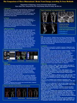
Radiology-To Scan or not to Scan? Dr Sue Rowe Consultant Radiologist HUH
Topics • Thyroid Ultrasound • MSK Imaging • CT KUB
Justification • IRMER • Also helps radiology do the correct test
Why Are You Requesting This Test? • What information are you hoping to gain? • Will this information alter patient management? • What information may you gain that creates anxiety?
Thyroid Ultrasound • Worldwide increase in thyroid ultrasound • GP demand stable at HUH but recent increase in requests with predicted rise of 35% this year (compared to 12-13).
CLINICAL HISTORY • Previous cancer • Immunocompromised • Previous radiation • Relevant previous operations/treatments • Imaging at another hospital • Relevant family history
Thyroid • Nearly 70% of the population have a thyroid nodule at ultrasound, 45% of this group>1 nodule • 52 million adults in the UK • 35 million nodules.. and some more follow ups… and some FNA’s • 87% increase in papillary cancer in USA is nodule <2cm and 49% <1cm.
In the USA • 2.4 fold increase in reported incidence of thyroid nodules over the last 30 years due to ultrasound. • 3 fold increase in FNA
Thyroid Cancer • Incidence of and mortality from thyroid cancer in the US, 1975-2009 and advent of new technologies Fine needle aspiration of thyroid nodules. Increase in use of CT and MRI Ultrasound guided aspiration of thyroid nodules. BMJ 2013; (347):18-21.
The incidence of thyroid cancer for men and women in England approximately doubled between 1990-94 and 2006-10.
Overdiagnosis Potential Harm • Unnecessary anxiety due to follow up ultrasounds, FNA’s, OPD’s • Potential ‘unnecessary’ thyroidectomy if inadequate FNA or papillary cancer which may have not progressed.
How Do We Avoid This? • Do not ultrasound purely for over or underactive thyroid • Ultrasound if palpable lump
Effective Dose in mSv (days of background radiation) • CXR 0.02 (1 day) • AXR 0.7 (50 days) • T Spine 1 (35 days) • L Spine 1.5 (65 days) • Knee 0.005 (< 1 day)
6mSv background radiation a year in Cornwall or the Scottish Highlands
Typical lifetime total cancer risk as a function of age at exposure and sex HPA-CRCE-028
MSK Appendicular • Axial-plain x-ray first. Better for ossification, foreign bodies, loose bodies, essential in primary tumour assessment • X-ray may guide whether you want to undertake MR or refer • Lumps and Bumps- ?nothing or ultrasound.
Shoulders • X-ray-useful for most conditions • Ultrasound- tendinopathy, injection, bursitis • Specialist referral if for dislocation
Knee • X-ray- severe degenerative change, don’t need MR • Age >50 very unlikely to warrant MR, unless ?fracture, infection, severe ligamentous injury or synovitis. Specialist referral/locomotive service. • Don’t repair menisci after age 40
Knee Ultrasound • Injection • Patellar and quadriceps tendon • ?Popliteal cyst • Pes Anserinus Bursa • Chronic MCL pain (stripping) • Iliotibial Band Friction • Muscle tears (aspiration)
Acute Trauma Knee • In the young patient serious concern regarding mensical tear or ligamentous injury consider referring urgently to sports clinic (Tues and Fri 020 8510 7835) . Small tear may increase in size if not treated urgently. • Older patients more likely to be treated conservatively
Ankle • X-Ray: trauma, degenerative change, inflammation • Ligamentous injuries may be initially treated conservatively, so don’t necessarily need definitive diagnosis. • MR if worried re fracture which has not been identified on x-ray. Should be able to weight bear without pain 1 month into rehab • Tender over talar dome ?OCD
Muscle tears in calf- if collection needs aspiration to facilitate healing • Fractures different rehab, especially if sports person • Late referral/diagnosis can immensely prolong rehab
Back Pain • Back pain affects most adults at some time, ? 20% of these consult their GP • 85-98% Non specific back pain • 70% back to work in 1 week, 90% within 2 months • Radicular Pain • Red Flags • Not MSK in origin (renal, GI, aorta, gynae)
Red Flags • Symptoms of Cauda Equina (sphincters, gait saddle anaesthesia) • Motor Loss • Elevated plasma viscosity • Systemic symptoms (weight loss, pyrexia) • Increased risk of TB or recent sepsis • Non mechanical back pain • Thoracic Pain (2/3rds mets in thoracic region) • Age <16 (cancer), 16-20 (seroneg arthropathy) or new pain >50 (cancer) • Previous cancer
X-Rays • Trauma or osteoporotic fracture • Don’t relay on findings. Any Red Flags consider specialist referral or MR; need 50% bone loss to see a lesion on x-ray. Path tests. • Seronegative arthritis will see late changes, again can’t exclude. Does not assess activity.
Stratification with STarT Back • Low Risk: self management with GP support • Medium risk: physio referral, exercise, manual therapy, TENS • High Risk: biopsychosocial assessment and management plan Maps of Medicine April 2013
Lower Back Pain • Utilise Locomotive Service if 4 weeks of intractable back pain • 6 weeks of conservative active management and still pain undertake MR
Specialist Referral • Radicular Pain- shooting or lancing pain with tingling, burning numbness in distribution of a nerve root • Severe Radicular Pain: disabling pain, intrusive, stops patient going to work • Neurological Deficit
NICE Guidance 2008 • Physical activity and exercise • Manual therapy [1] • Invasive procedures • Combined physical and psychological treatment programme • Consider referral for a combined physical and psychological treatment programme • Assessment and imaging • Do not offer X-ray of the lumbar spine for the management of non-specific low back pain. • Only offer an MRI scan for non-specific low back pain within the context of a referral for an opinion on spinal fusion (see section 1.9). • Referral for surgery • Consider referral for an opinion on spinal fusion for people who: have completed an optimal package of care, including a combined physical and psychological treatment programme (see section 1.7) and still have severe non-specific low back pain for which they would consider surgery.
Reassurance? • Patients may develop a sense of ‘less well being’ when the extent of their spine disease is revealed • Around 1/3rd of radiculopathy symptomatic discs will regress at 6 weeks • Increased frequency of lumbar spine MR associated with higher rates of spinal surgery without clear differences in patient outcomes • Radiology 2005;237:597-604 • Mapsofmedicine.com
Lumbar Canal Stenosis • No correlation between imaging appearances and the level of disability • MR used to guide surgery levels • Eur Spine J. 2008 May; 17(5): 679–685.
Neck Pain • Common condition, up to 70% adults • Acute torticollis • Whiplash • Non-specific neck pain • Cervical radiculopathy • Others • Red flags
Red Flags • Trauma X-Ray • Inability to rotate the neck 45 degrees • Focal neurological deficit/ weakness • Dangerous mechanism of injury • Myelopathy • Cancer, infection etc
X-Ray • Indicated in trauma. • Red Flags, but don’t use to exclude • Rheumatoid Arthritis and seronegative arthropathies
MRI • Red flags • Consider if failure to respond to treatments over (?)12 weeks • Radiculopathy- or refer?
Informally run for several years Should follow ELIC protocol Not a substitute for referral to haematuria clinic Do requests for CT KUB meet the agreed protocol? What is the positive rate for renal colic CT KUB
CT KUB • 46 CT KUB over the period Dec 2012 to May 2013 • Request details and reports evaluated • 30 pts haematuria, 1 no haematuria, 15 not stated • 2 patients macroscopic haematuria, 13 micro, not stated 31 • Temperature only mentioned in 1 • 10 no side of pain mentioned • 6 (13%) positive for renal colic, 9 (20%) renal calcifications, no significant other findings • 2009 Audit ED referrals 48.5% renal colic, 9.5% significant other findings