Download
1 / 33
330 likes | 435 Views
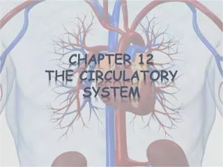
Chapter 12 The Circulatory System. HEART. Location, size, and position Triangular organ located in mediastinum two thirds of the mass to the left of the body midline and one third to the right the apex on the diaphragm shape and size of a closed fist (Figure 12-1). CPR.

E N D
HEART • Location, size, and position • Triangular organ • located in mediastinum • two thirds of the mass to the left of the body midline and one third to the right • the apex on the diaphragm • shape and size of a closed fist (Figure 12-1)
CPR • Cardiopulmonary resuscitation (CPR)— • the heart lies between the sternum in front and the bodies of the thoracic vertebrae behind • rhythmic compression of the heart between the sternum and vertebrae can maintain blood flow during cardiac arrest • http://medicine.arizona.edu/spotlight/learn-sarver-heart-centers-continuous-chest-compression-cpr
HEART • Anatomy • Heart chambers (Figure 12-2) • Two upper chambers called atria (receiving chambers)—right and left atria • Two lower chambers called ventricles (discharging chambers)—right and left ventricles • Wall of each heart chamber is composed of cardiac muscle tissue called myocardium • Endocardium—smooth lining of heart chambers—inflammation of endocardium called endocarditis
Heart • Anatomy-con’t • Covering sac, or pericardium- • Pericardium is a two-layered fibrous sac with a lubricated space between the two layers • Inner layer called visceral pericardium or epicardium • Outer layer called parietal pericardium
HEART • Anatomy • Heart action • Contraction of the heart is called systole • Relaxation is called diastole
Heart valves (Figure 12-3) • Valves keep blood flowing through the heart and prevent backflow • Consist of two atrioventricular, or AV, and two semilunar (SL) valves • Tricuspid—at the opening of the right atrium into the ventricle • Bicuspid (mitral)—at the opening of the left atrium into the ventricle • Pulmonary semilunar—at the beginning of the pulmonary artery • Aortic semilunar—at the beginning of the aorta
BLOOD VESSELS • Types • Arteries—carry blood away from the heart • Veins—carry blood toward the heart • Capillaries—carry blood from the arterioles to the venules
BLOOD VESSELS • Structure (Figure 12-9) • Arteries • Tunica intima—inner layer of endothelial cells • Tunica media—smooth muscle with some elastic tissue, thick in arteries; important in blood pressure regulation • Tunica externa—thin layer of fibrous elastic connective tissue
Capillaries—microscopic vessels with only one layer—tunica intima • Veins • Tunica intima—inner layer; valves prevent retrograde movement of blood • Tunica media—smooth muscle; thin in veins • Tunica externa—heavy layer of fibrous connective tissue in many veins
BLOOD VESSELS • Functions • Arteries—distribution of nutrients, gases, etc., with movement of blood under high pressure; assist in maintaining the arterial blood pressure • Capillaries—serve as exchange vessels for nutrients, wastes, and fluids • Veins—collect blood for return to the heart; low pressure vessels
Names of main arteries—see Figure 12-10 and Table 12-1 • Names of main veins—see Figures 12-11 and 12-12 and Table 12-2
HEART • Blood supply to the heart muscle • Blood, which supplies oxygen and nutrients to the myocardium of the heart, flows through the right and left coronary arteries (Figure 12-5); called coronary circulation • Blockage of blood flow through the coronary arteries is called myocardial infarction (heart attack)
Angina pectoris—chest pain caused by inadequate oxygen to the heart • Coronary bypass surgery—veins from other parts of the body are used to bypass blockages in coronary arteries (Figure 12-6)
CIRCULATION • Systemic and pulmonary circulation Refers to the blood flow through the vessels arranged to form a circuit or circular pattern (Figure 12-13)
Pulmonary circulation • Carries blood to and from the lungs; arteries deliver deoxygenated blood to the lungs for gas exchange • Path goes from right ventricle through pulmonary arteries, lungs, pulmonary veins, to left atrium
Systemic circulation • Carries blood throughout the body • Path goes from left ventricle through aorta, smaller arteries, arterioles, capillaries, venules, venaecavae, to right atrium
HEART • Blood flow through the heart (Figure 12-4) • The heart acts as two separate pumps—the right atrium and ventricle performing different functions from the left atrium and ventricle
Pulmonary Circulation • venous blood enters the right atrium through the superior and inferior venae cavae • passes from the right atrium through • the tricuspid valve to the right ventricle; • from the right ventricle it passes through the pulmonary semilunar valve to the pulmonary artery to the lungs
Systemic Circulation • Blood moves from the lungs to the pulmonary veins into the left atrium, • passing through the bicuspid (mitral) valve to the left ventricle; • blood in the left ventricle is pumped through the aortic • semilunar valve • into the aorta and is distributed to the body as a whole
CIRCULATION • Hepatic portal circulation (Figure 12-14) • Unique blood route through the liver • Vein (hepatic portal vein) exists between two capillary beds • Assists with homeostasis of blood glucose levels
CIRCULATION • Fetal circulation (Figure 12-15) • Refers to circulation before birth • Modifications required for fetus to efficiently secure oxygen and nutrients from the maternal blood • Unique structures include the placenta, umbilical arteries and vein, ductus venosus, ductus arteriosus, and foramen ovale
Next week • Unit 3 Seminar • Chapter 13 • The Lymphatic System and Immunity