Download
1 / 32
320 likes | 527 Views
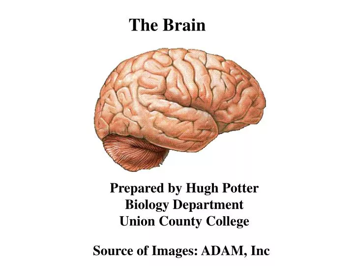
The Brain . Prepared by Hugh Potter Biology Department Union County College. Source of Images: ADAM, Inc. BRAIN - LATERAL VIEW. COVERINGS OF THE BRAIN. A. B. A. THE CRANIUM B. THE MENINGES. The Meninges

E N D
The Brain Prepared by Hugh PotterBiology DepartmentUnion County College Source of Images: ADAM, Inc
COVERINGS OF THE BRAIN A B A. THE CRANIUM B. THE MENINGES
The Meninges The Meninges are tough, moist membranes surrounding the brain and spinal cord. Infection of meninges is called meningitis. 1. Dura mater – outermost membrane. Very tough, protective layer consisting of: a. The Periosteal layer – thick, outer portion, continuous with the periosteum of the cranial bone. b. Meningeal layer- thin, inner portion. The spinal dura mater consists only of this layer. 2. Arachnoid layer – a fibrous, delicate middle zone. Below this layer is the subarachnoid space filled with cerebrospinal fluid. 3. Pia mater – innermost of the meningeal layers. It is closely adherent to the surface of the brain and spinal cord.
Cerebrospinal Fluid and the Ventricles of the Brain CSF is manufactured by specialized blood vessels, the choroid plexi. These vessels are located within spaces of the brain called the ventricles. The brain contains four ventricles: The Lateral ventricles – Each cerebral hemisphere contains a lateral ventricle (ventricles I and II). They are separated from one another by a thin partition called the septum pellucidum. The Third Ventricle – This ventricle is found below the lateral ventricles in the diencephalon. Each lateral ventricle is connected to ventricle III by a narrow opening – the Foramen of Monro. The Fourth Ventricle – This ventricle is found between the brain stem and the cerebellum. The III and IV ventricles are connected by a channel called the Aqueduct of Sylvius. The floor of the IV ventricle has three small holes which allow the CSF to leave the inside of the brain and pass into the subarachnoid space
The Cerebral Cortex The cerebral cortex is a structure within the brain that plays a key role in memory, attention, perceptual awareness, thought, language, and consciousness. In non-living, preserved brains, the outermost layer of the cerebrum has a gray color, hence the name gray matter. Gray matter is formed by neurons and their unmyelinated fibers, whereas the white matter below the gray matter of the cortex is formed predominantly by myelinated axons interconnecting different regions of the central nervous system. The human cerebral cortex is 2-4 mm (0.08-0.16 inches) thick. The surface of the cerebral cortex is folded in large mammals, wherein more than two-thirds of the cortical surface is buried in the grooves, called “sulci."
Connections of the Cerebral Cortex to other Brain Regions The cerebral cortex is connected to various structures such as the thalamus and the basal ganglia, sending information to them along efferent connections and receiving information from them through afferent connections. Most sensory information is routed to the cerebral cortex from the thalamus. Olfactory information, however, passes through the olfactory bulb to the olfactory cortex directly. The vast majority of connections are from one area of the cortex to another rather than to subcortical areas. The cortex is commonly described as comprising three parts: sensory, motor, and association areas.
Sensory areas The sensory areas are the areas that receive and process information from the senses. Parts of the cortex that receive sensory inputs from the thalamus are called primary sensory areas. The senses of vision, audition, and touch are served by the primary visual cortex, primary auditory cortex and primary somatosensory cortex, respectively. In general, the two hemispheres receive information from the opposite (contralateral) side of the body. For example the right primary somatosensory cortex receives information from the left limbs, and the right visual cortex receives information from the left visual field. Areas with lots of sensory innervation, such as the fingertips and the lips, require more cortical area to process finer sensation.
Association areas Association areas function to produce a meaningful perceptual experience of the world, enable us to interact effectively, and support abstract thinking and language. The parietal, temporal, and occipital lobes organize sensory information into a coherent perceptual model of our environment centered on our body image. The frontal lobe or prefrontal association complex is involved in planning actions and movement, as well as abstract thought. Our language abilities are localized to the association areas of the parietal-temporal-occipital complex, typically in the left hemisphere. Two of these areas are most important:1. Wernicke’s area relates to understanding the meaning of language, facial expressions and body language.2. Broca’s area controls the muscles of the tongue, lips and larynx that we use to produce speech.
Motor areas The motor areas are located in both hemispheres of the cortex. They are shaped like a pair of headphones stretching from ear to ear. The motor areas are very closely related to the control of voluntary movements, especially fine fragmented movements performed by the hand. The right half of the motor area controls the left side of the body, and vice versa. Two areas of the cortex are commonly referred to as motor: 1. Primary motor cortex, which executes voluntary movements 2. Premotor cortex, Regulates skilled motor activities. This area “instructs” the Primary Motor cortex.which then selects voluntary movements.
THE BRAIN - SAGITTAL VIEW A A. The Cerebrum B. The Cerebellum C. The Pons D. The Medulla Oblongata C B D
THE BRAIN - LATERAL VIEW I The Cerebrum A - Frontal Lobe B - Parietal Lobe C - Temporal Lobe D - Occipital LobeII The Cerebellum B A D C II
CEREBRUM - FRONTAL LOBE 1. The frontal lobe initiates motor activities. 2. It controls the movements of the tongue leading to speech through Broca’s area.3. The frontal lobe controls the formation of thought and emotions and stores memory. 4. The frontal lobe is the region from which our personality and intelligence originates.
CEREBRUM - PARIETAL LOBE The Parietal lobe receives sensory nerve information from various regions of the body, especially the skin. This is called general sensation. The parietal lobe is also responsible for interpreting this information
Cerebrum - Temporal Lobe 1. The temporal lobe is responsible for the perception and interpretation of auditory input. 2. An Olfactory Area receives and interprets odors. 3. Wernicke’s area allows us to put words to auditory, visual or even touch sensations (Braille). This area turns these sensations into meaningful ideas. Temporal lobe
CEREBRUM - OCCIPITAL LOBE The Occipital lobe contains the Visual Area responsible for the interpretation of visual input or sight. It is the destination of the optic nerve.
CORPUS CALLOSUM The Corpus callosum is a flat, sheet-like nerve tract connecting the right and left cerebral hemispheres. It contains more than 200 million axons.
Basal Cerebral Ganglia1. Nuclei buried deep in the cerebrum just superior to the thalamus. A nucleus is a group of nerve cell bodies found together in a specific area of the central nervous system 2. The basal ganglia control the background, gross, intentional body movements which are always associated with the more precise movements of arms, hands, fingers and feet. For example, when you are picking up a pencil, the conscious reach and grasp actions are performed by the arm, hand and fingers. The cerebral nuclei position your shoulder and stabilize the arm to make the grasp more precise. 3. The basal nuclei help maintain body posture and muscle tone.
Limbic System The limbic system is found along the border between the cerebrum and the diencephalon. The limbic system links the conscious functions of the cerebral cortex with the unconscious and autonomic functions of the lower brain. It plays a very large role in memory storage and retrieval. Temporal lobe hippocampus
Components of the Limbic System1. Hippocampus is a sea horse shaped region responsible for establishing new memories, especially those that are emotionally charged. 2. Mamillary body processes sensory information and movements associated with eating, e.g., chewing, licking and swallowing. 3.Amygdala is crucial in the formation of emotions. It is highly connected to our learning and memory centers. The amygdala is central to most brain events involving fear. 4. The Fornix is a tract of white matter that connects the hippocampus with the hypothalamus.
The hippocampus is located in the medial portion of the temporal lobe of the cerebrum. It is part of a nerve tract called the limbic system. The name is based on its shape – like a sea horse.
Why should you care about the hippocampus? Here are three good reasons. First, this part of the brain appears to be absolutely necessary for making new memories. If you didn't have it, you couldn't live in the present: you'd be stuck in the past of old memories. Alzheimer's disease affects the hippocampus first and very severely, before other parts of the cortex (later, the frontal lobes too). So memory is usually the first thing to start to falter in Alzheimer's -- the ability to make new ones, that is. Who visited yesterday? Where did I put the car keys? Why isn't there any mail today (when you brought it in 3 hours ago)?
Secondly, the hippocampus seems to be involved in severe mental illnesses. In both schizophrenia and some severe depressions, the hippocampus appears toshrink. However, there is recent evidence that this shrinkage can be reversed and perhaps prevented in people with depression and bipolar disorder, with effective treatment.
Third, the hippocampus is known to be directly affected by estrogen. Estrogen increases "synaptic density" -- the number of connections to other nerve cells -- in the hippocampus. There is some evidence that estrogen may play a role in preventing Alzheimer's.
The Diencephalon • The diencephalon is located directly under the corpus callosum. The third ventricle is found in the diencephalon. The diencephalon is composed of three major regions: • TheThalamus is a motor relay station for sensory information from the spinal cord to the cerebral cortex. All sensory information going to the brain (except for olfactory) has to make a pit stop in the thalamus. • The Epithalamus is located in the most superior and anterior portion of the diencephalon. The choroid plexus of the third ventricle is found in the epithalamus. The pineal body is also found here. The pineal manufactures melatonin which helps to regualte our sleep-wake cycle. • The Hypothalamus is located just below the thalamus on the floor of the brain. It is a major center for the control of internal body functions through the activities of several nuclei.
Regulatory Centers of the Hypothalamus 1. Preoptic nucleus – controls body temperature. 2. Supraoptic nucleus - regulates the secretion of anti-diuretic hormone. This hormone helps to regulate the balance between fluids and electrolytes in the body (osmoregulation), as well as, blood pressure. 3. Paraventricular nucleus – manufactures the hormone oxytocin which stimulates the smooth muscle of the uterus. 4. Medial nuclei – produce a sense of satiety or satisfaction from food intake due to a rise in blood sugar. 5. Lateral regions of the hypothalamus – Stimulation of this area produces the feeling of hunger of thirst depending on the precise location stimulated. 6. The hypothalamus exerts a great deal of control over the function of the pituitary gland.
CEREBELLUM The cerebellum is located posterior to the pons and medulla oblongata. Through its nerve connections to the cerebrum and spinal cord, it coordinates all motor activity of the body.
The Brain Stem The brain stem is the last major portion of the brain. It contains the centers for regulating physiological processes, e.g., respiration, blood pressure, heart rate and wake-sleep cycles. Midbrain – is the uppermost region of the brain stem. The Pons – is located just superior to the medulla oblongata and inferior to the midbrain. Medulla oblongata – is the last region of the brain stem before the spinal cord.
The Midbrain • the uppermost region of the brain stem. • 2. located at the junction of the cerebellum and pons. • 3. a relay station for motor and sensory messages between • parts of the brain and the spinal cord. • 4. The tectum of the midbrain contains two pairs of • rounded bulges, the corpora quadrigemina. The • superior colliculi coordinate motor reflexes produced by • visual stimuli. The two inferior colliculi coordinate motor • reflexes due to auditory input. • 5. The substantia negra is located in the midbrain. The substantia • negra controls motor tracts form the cerebrum by releasing an • inhibitory neurotransmitter, dopamine. Damage to this area leads to • Parkinson’s disease.
The Pons The Pons is located just superior to the medulla oblongata and inferior to the midbrain. It contains many of the longitudinal nerve tracts passing through the brain stem: 1. Corticospinal tracts – The axons of these tracts originate at the cerebral cortex and pass directly to the motor neurons of the spinal cord. 2. Spinothalamic tracts – are sensory tracts carrying nerve messages of crude touch, pain and temperature from various levels of the spinal cord to the thalamus and then to the cerebral cortex. The pons contains nuclei for regulating respiratory movements: 1. The apneustic center stimulates the respiratory rhythmicity center (RRC) in the medulla oblongata to produce inspiration. 2. The pneumotaxic center inhibits the RRC producing a passive expiration.
The Medulla Oblongata The medulla oblongata is the last portion of the brain before the spinal cord. The medulla contains the corticospinal and spinothalamic nerve tracts. 1. Corticospinal tracts contain descending nerve fibers for the control of conscious motor activity. In the lower medulla, these tracts decussate (cross over). As a result, the left cerebral cortex initiates motor activity on the right side of the body and vice versa. 85% of the corticospinal tracts decussate. 2. The Spinothalamic tracts pass through the medulla and converges on the thalamus. They carry sensory information of crude touch, pressure and pain, as well as, thermal sensation. There is no decussation of these messages in the medulla. This has already occurred in the spinal cord at the level of entry to the medulla.
Medullary Nuclei Medullary nuclei are clusters of cell bodies within the interior of the medulla oblongata that carry out a variety of regulatory functions: 1. Cardiovascular centers regulate heart rate, and vasomotion. 2. Respiratory rhythmicity center sets the breathing pace with input from the apneustic and pneumotaxic centers of the pons.