Acknowledgements
Step 3. Step 4. Figure 3 . Steps #3-7 for computing local cortical thickness. Step 5 Step 6 Step 7. Step 1. Step 2. Step 3.

Acknowledgements
E N D
Presentation Transcript
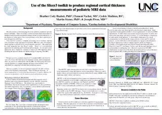
Step 3. Step 4 Figure 3. Steps #3-7 for computing local cortical thickness. Step 5 Step 6 Step 7 Step 1. Step 2. Step 3. Use of the Slicer3 toolkit to produce regional cortical thickness measurements of pediatric MRI data Heather Cody Hazlett, PhD1; Clement Vachet, MS1; Cedric Mathieu, BS1; Martin Styner, PhD2; & Joseph Piven, MD1,3 1Department of Psychiatry, 2Department of Computer Science, 3Carolina Institute for Developmental Disabilities Background The data analysis of neuroimaging data from pediatric populations presents several challenges. There are normal variations in brain shape from infancy to adulthood and normal developmental changes related to tissue maturation (i.e., myelination of white matter) that create problems in the direct application of tools designed for adult brain. Our team has created a computer processing tool to produce regional cortical thickness maps appropriate for pediatric MRI data, and is developing a similar pipeline to perform local cortical thickness measures. This application has been integrated into the Slicer3 toolkit. Slicer3 is a cross-platform application for analyzing and visualizing medical images. It is an open source application and is funded by a number of large-scale NIH supported efforts, including the National Alliance for Medical Image Computing (NA-MIC). Local cortical thickness requires a mesh-based methodology, which involves many more steps than regional cortical thickness applications. Main components for this pipeline include (1) tissue segmentation, (2) atlas-based ROI segmentation, (3) white matter map creation and post-processing, (4) genus-zero white matter map image and surface creation, (5) cortical thickness computation, (6) white matter mesh inflation, (7) sulcal depth computation, (8) cortical correspondence on inflated meshes using a particle system, and (9) statistical analysis. As described above in the description of regional cortical thickness, the Slicer3 application is able to compute steps #1 and #2 (see Figure 1). Currently, several applications have been developed and integrated within Slicer3 for steps #3-7 (see Figure 3 below), and the last step regarding cortical correspondence is undergoing tests. However, as part of our NA-MIC collaboration, we plan to have the whole the mesh-based local cortical thickness analysis pipeline fully working and integrated within Slicer3. these steps if the related images are provided, such as tissue segmentation label maps or parcellation maps. Figure 2. Pipeline steps involved to produce regional cortical thickness measures. Methods We have access to a pediatric dataset of T1-weighted MRI scans from 90 cases of 2-4 year olds with typical development, autism, and developmental delay. As part of our collaboration with NA-MIC our UNC group will create separate pipelines to compute regional and local cortical thickness, and algorithms for computing both individual and group analysis. There are a number of critical components required to be able to obtain valid cortical thickness measurements. These include the following: (1) tissue segmentation, (2) cortical thickness measures, (3) surface inflation, (4) cortical correspondence, and (5) statistical analysis/hypothesis testing. REGIONAL CORTICAL THICKNESS A Slicer3 high-level module performing individual regional cortical thickness analysis has been developed by our lab: ARCTIC (Automatic Regional Cortical ThICkness). The default basic steps include (1) probabilistic atlas-based automatic tissue segmentation, (2) atlas parcellation deformable registration, and (3) asymmetric cortical thickness measurement. See example of these steps below (Figure 1): The ARCTIC application provides not only lobar cortical thickness but also tissue segmentation volume information stored in spreadsheets. Moreover a quick quality control can be performed for each step within Slicer3 using a MRML scene displaying output volumes and surfaces. ARCTIC is still in development in order to improve its integration within Slicer3, but upon completion of our NA-MIC collaborative project we plan to have ARCTIC available cross-platform, with Windows and MAC executables available on NITRIC. Plans are also underway to make ARCTIC’s source code publically available via a SVN repository. Regarding validity tests, ARCTIC has been tested on our pediatric dataset. Results are available on the NA-MIC wiki page. Current work is underway to conduct validity tests against other applications (e.g., FreeSurfer). In progress is a test using 40+ cases from FreeSurfer’s publically available tutorial dataset. Future Directions Current work in our lab is underway to develop a pipeline for local cortical thickness. With this application, we will be able to generate cortical thickness maps across the entire surface of the brain and compute group-based comparisons in cortical thickness measurements. Acknowledgements Research supported by NIMH grants MH61696 and MH64580 (PI: Joseph Piven, MD) and PHS-NIH EB005149-04 (PI: Kikinis) National Alliance for Medical Image Computing (NA-MIC). Resources Available to the Public All documentation for this application and the Slicer3 toolkit is available on the NAMIC wiki pages, including two tutorials describing the cortical thickness applications developed by our UNC group. (http://www.na-mic.org). ARCTIC’s first release is publicly available on the NITRC website (http://www.nitrc.org/projects/arctic). Pediatric and adult brain atlases used by ARCTIC are also available on MIDAS (http://www.insight-journal.org/midas/collection/view/34). The next figure (Fig. 2) provides a summary graphic detailing the main components of the regional pipeline required to generate the regional cortical thickness of the dataset. The user has the possibility to skip some of