Download
1 / 38
1.36k likes | 5.51k Views
VP Shunts. Division of Child Neurology Department of Pediatrics Goryeb Children’s Hospital Atlantic Health System. Cerebral Shunts. To treat hydrocephalus / reduce ICP Proximal end inserted into a CSF source (usually blocked) Ventricle Lumbar cistern of the spinal cord

E N D
VP Shunts Division of Child Neurology Department of Pediatrics Goryeb Children’s Hospital Atlantic Health System
Cerebral Shunts • To treat hydrocephalus / reduce ICP • Proximal end inserted into a CSF source (usually blocked) • Ventricle • Lumbar cistern of the spinal cord • Distal end inserted near absorptive epithelial surface or directly into the blood stream: • Peritoneal cavity of the abdomen (most common) • VP shunt = ventriculo-peritoneal shunt • LP shunt = lumbar-peritoneal shunt • Right Atrium of the heart (VA shunt) • Pleural cavity of the lung (VPL shunt)
VP SHUNT VA SHUNT
Cerebral Shunts • May also insert distal end into: • gallbladder (mixes with bile) • ureter (mixes with urine) • Variety of forms: • made of different materials (silicone) • different types of pumps and uni-directional valves • +/- programmable
Shunt Complications • More common in childhood • May require immediate shunt revision or shunt re-programming • Shunt complications often mimic the symptoms that prompted initial shunting • headache • double vision • nausea / vomiting • altered mentation (lethargy / irritability) • bulging fontanelle • Shunt failure rate 2 years after insertion - up to 50%
“Sunsetting Eyes”: clinical sign of increased intracranial pressure
Infection • Incidence 1-20 %, average 10 % • Usually intra-operative contamination of surgical wound by skin flora • Common microbial agents • Staph epi (coagulase negative staph) > 50% • Staph aureus 20 % • Gram negative bacilli 15 % • Candida • Symptoms – ICP, fever, WBC • No correlation with shunt type • Risk factors for shunt infection • age < 6 months
4 Distinct Clinical Syndromes of Shunt Infection 1. Colonization of the shunt - most common 2. Wound infection 3. Peritonitis / distal infection 4. Meningitis
1. Colonization of the Shunt • MOST COMMON • Symptoms of shunt malfunction > infection • Lethargy, headache, vomiting, full fontanelle • Low grade fever • Within months of shunt insertion • CSF from ventricle or lumbar puncture STERILE • Unusual to see signs of meningitis / ventriculitis • CSF minimally abnormal • Infecting organism in SHUNT RESERVOIR • Blood cultures negative unless VA Shunt colonization
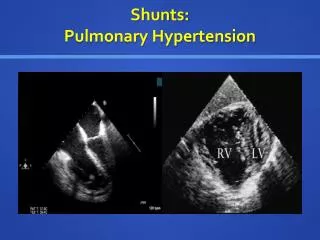
If VA shunt, more severe systemic symptoms • Septic pulmonary emboli • Pulmonary hypertension • Infective endocarditis • For more chronicVA shunt colonization • hypo-complementemic glomerulonephritis = Ag-Ab complex deposition in glomeruli • “Shunt Nephritis” • hypertension, microscopic hematuria, elevated BUN and creatinine, anemia
2. Wound Infection • Obvious infection or dehiscence along the shunt tract • Within days-to-weeks of shunt procedure • Staph aureus - most common isolate • Fever common • Symptoms of shunt malfunction follow
3. Distal Infection / Peritonitis • Abdominal symptoms without signs of shunt malfunction common • Pathogenesis: • perforation of bowel at time of insertion • translocation of bacteria across the bowel wall • Gram negative isolates, mixed flora cultured from distal portion of shunt catheter
4. Meningitis • Pathogens: • Strep pneumo • N. meningitidis • Hib • Presentation typical of acute bacterial meningitis
Treatment of Shunt Infection 1. IV anti-staph PCN (if resistant, IV vancomycin) 2. intra-shunt vancomycin (monitoring CSF levels to avoid toxicity) • due to poor penetration of most abx into CSF across inflamed meninges 3. externalize the distal shunt For gram negative infections : • 3rd generation IV cephalosporin • Intra-shuntaminoglycoside
Treatment of Shunt Infection • Often need to remove shunt • colonization, wound infection, distal peritonitis • for meningitis, IV abx without shunt removal • After reservoir CSF sterile x 48 hour, can insert new shunt on other side • High rate of infection relapse due to: • Abx therapy alone (no shunt externalization or removal) • Abx therapy + partial shunt revision
Prevention of Shunt Infection • Meticulous cutaneous preparation and surgical technique • ?? perioperative IV abx, intra-ventricular abx, abx impregnated shunt tubing, soaking the shunt in abx
Other Shunt Complications • Obstruction • Proximal – build-up of excess protein in CSF, slowly clogs the valve • Distal – build-up of excess peritoneal protein blocks distal tip • Over-drainage (see below) • Slit Ventricle Syndrome (see below) • Over-drainage • Intraventricular CSF drains too rapidly brain collapses on itself extra-axial fluid (CSF or blood) collects to fill the spatial void external compression of brain brain damage, stretching of bridging veins subdural hemorrhage
Other Shunt Complications • Slit Ventricle Syndrome • CSF slowlyover-drains over several years after shunt procedure • uncommon, but results in need for many shunt revisions • symptoms similar to typical shunt malfunction BUT • cyclical (appear, subside, appear, subside…, over years) • symptoms alleviated by lying prone • due to: • overdrainage simultaneous with brain growth (brain growth fills the intraventricular space, leaving the ventricles collapsed) • compliance of brain decreases, preventing ventricles from enlarging • collapsed ventricles can also block shunt valve (a form of obstruction)
Slit Ventricle • Syndrome
Other Shunt Complications • Intra-ventricular hemorrhage • occurs at any time during or after a shunt insertion or revision • can occur in nearly 31% of shunt revisions
A large dural hole around the ventricular catheter may predispose to CSF flow through the dural opening leading to the formation of subcutaneous tract
Obstructive / Non-communicating Hydrocephalus due to Aqueductal Stenosis • CT of the brain: • large frontal and • temporal horns of • lateral ventricles • large third ventricle • 4th ventricle small 3rd 4th
Obstructive / Non-communicating Hydrocephalus due to Chiari Malformation low lying tonsils alone (Chiari I) – usually asymptomatic low lying tonsils + hydrocephalus (Chiari II) – diffuse headache Chiari I Chiari II (+ lumbosacral myelomeningocele)
Non-Obstructive / Communicating Hydrocephalus as a complication of prior Meningitis CT of the brain reveals enlarged frontal and temporal horns of the lateral ventricles and enlarged 3rd and 4th ventricles. 3rd 4th
Dandy-Walker Malformation: aplasia / hypoplasia of cerebellar vermis (midline cerebellum missing or underdeveloped)
Hydrocephalus due to Choroid Plexus Papilloma (CSF secreting intraventricular tumor which obstructs ventricular system)
Conditions with enlarged CSF spaces that usually do NOT require shunting
Lissencephaly “smooth brain” - achieve maximum 3-5 month dev milestones - may be caused by LIS-1 gene mutation (Miller-Diecker lissencephaly) - microcephaly, MR, seizures
Multifocal Cystic Encephalomalacia (hx of neonatal meningitis)