Download
1 / 1
10 likes | 84 Views
Study on Survivin and XIAP expression in renal cell carcinoma metastases to lungs, with correlations to clinical parameters and survival rates, suggesting potential implications for metastasis processes.

E N D
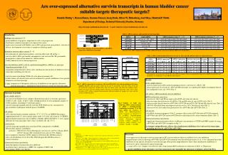
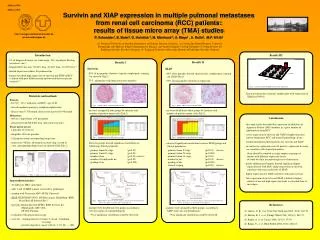
Survivin and XIAP expression in multiple pulmonal metastases from renal cell carcinoma (RCC) patients: results of tissue micro array (TMA) studies P. Schneider1, B. Baier2, O. Holotiuk 3, M. Meinhard 4, A. Meye1 , A. Rolle2, M.P. Wirth1 http://urologie.uniklinikum-dresden.de ptr.schneider@gmx.de Introduction Results II Results I • 3% of diagnosed tumors are renal origin, 50% of patients develop • metastases (ms) 1 • - frequent RCC ms-sites: 50-60% lung, 30-40% bone, 30-35% liver 2 • - limited expression studies for pulmonal ms • - former described high expression of survivin and XIAP in RCC • is linked with poor differentiation and lowered disease specific • survival 3,4 Survivin XIAP - 89% of ms punches showed a typical cytoplasmatic staining for survivin (Fig.2) - 71% of punches with lung tissue were negative - 80% of ms punches showed characteristic cytoplasmatic staining for XIAP (Fig.5) - 93% of lung punches showed no expression Fig.6 Coherency between XIAP- and survivin-expression 4x 40x 4x 40x Fig.2 Punch of RCC stained with anti-survivin Fig.4 Punch of RCC stained with anti-XIAP - ms were arranged in four groups by intensity and number of positive tumor cells (Tab.1) - ms were divided into three groups by intensity and number of positive tumor cells (Tab.2) Tab.1 Arrangement of ms, allocation to groups Tab.2 Arrangement of ms, allocation to groups - Survivin-groups showed significant correlations to following clinical parameters - observed significant correlations between XIAP-groups and clinical parameters - primary tumor N-stage (p<0.01) - primary tumor G-stage (p<0.01) - number of ms (p<0.01) (invers) - number of lymph node ms (p<0.05) - grading of ms (p<0.001) - primary tumor T-stage (p<0.01) (invers) - primary tumor N-stage (p<0.001) - primary tumor G-stage (p<0.01) - number of ms (p<0.01) (invers) - grading of ms (p<0.001) - clinical staging (p<0.05) (invers) Fig.3 Kaplan-Meier-curves for disease-time (a), disease-free-intervall (b) and time to ms-relapse (c) with results of log-rank-test Fig.5 Kaplan-Meier-curves for disease-time (a), disease-free-intervall (b) and time to ms-relapse (c) with result of log-rank-test - patients were divided into two groups according to survivin-status of corresponding ms - patients were arranged in three groups, according to XIAP-status of corresponding ms no significant correlations could be observed Abstract Nr: Abstract Id: (1) Technical University of Dresden, Department of Urology, Dresden, Germany, (2) Coswig Specialised Hospital, Center for Pneumology and Thoracic Surgery, Department of Thoracic and Vascular Surgery, Coswig, Germany, (3) Group Practice for Pathology Dresden, Dresden, Germany, (4) Technical University of Dresden, Institute of Pathology, Dresden, Germany Results III - Survivin expression correlates significantly with expression of XIAP (p<0.0001) Materials and methods Patients - 84 (52♂, 32♀) with clear cell RCC, age 43-82 - all with nephron-sparing or complete nephrectomy - disease-time 5-376 month, disease-free-interval 0-348 month Metastases - 606 ms, ranged from 1-64 per patient - all resected with Nd:YAG-laser (precision resection) Tissue micro arrays - 2 punches of every ms - altogether 1201 ms-punches - 118 punches from corresponding lung tissue - arrayed on 9 blocks, first punch on outer ring, second at core, corresponding lung-tissue on separate field (Fig.1) Conclusions - this study firstly described the expression of Inhibitors of Apoptosis Protein (IAP) members in a great number of pulmonal ms from RCC - (over)-expression of survivin and XIAP is higher than data known for primary RCC and normal kidney/lung- tissue - marked intratumoral heterogeneity for survivin and XIAP - in contrast to expression-score of patients, expression in single ms correlates with clinical parameters ms should be counted as single tumors or groups of tumors with different expression levels could be a hint for repeated process of metastasis - poorly differentiated tumors showed significant higher expression for both IAPs, higher expression of survivin correlates with increased levels of XIAP - higher expression for XIAP correlates with poor survival - low expression of survivin and XIAP is linked to higher number of ms and high expression leads to extended time to ms-relapse a b c Fig.1 Configuration of TMA (a), H&E stained 4µm slide (b), H&E stained punch (c) Immunohistochemistry - 10 slides per TMA, 4µm thick - slide 1 and 10 H&E stained, reviewed by pathologist - staining with Vectastain ABC-AP Kit Universal - XIAP: hILP/XIAP 610762, BD Biosciences, Heidelberg, BRD Vector Red AP Substrat Kit 1 - Survivin: human Survivin AF886, R&D Systems Inc., Minneapolis, MN, USA AP-New-Fuchsin a b a b c c References (1) Janzen, N. K. et al. Urol Clin North Am 2003; 30(4): 843-52. (2) Motzer, R. J. et al. N Engl J Med 1996; 335(12): 865-75. (3) Parker, A. et al. Cancer 2006; 107(1): 37-45. (4) Ramp, U., et al. Hum Pathol 2004; 35(8): 1022-8. • - evaluation with optical microscope • score for: staining intensity (0=none, 1=weak, 2=medium, • 3=strong) • percent of positive cancer-cells (0, 5, 10, 20,…, 100) no significant correlations could be observed