Download
1 / 1
10 likes | 202 Views
ELASTICITY IMAGING OF THE BREAST: A CLININCAL PERSPECTIVE Joseph R. Grajo BS 1 , Richard G. Barr MD, PhD 1,2 1 Northeastern Ohio Universities College of Medicine, Rootstown OH 2 Radiology Consultants, Inc., Youngstown OH. BACKGROUND. MATERIALS AND METHODS.

E N D
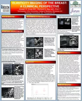
ELASTICITY IMAGING OF THE BREAST: A CLININCAL PERSPECTIVE Joseph R. Grajo BS1, Richard G. Barr MD, PhD1,2 1Northeastern Ohio Universities College of Medicine, Rootstown OH 2Radiology Consultants, Inc., Youngstown OH BACKGROUND MATERIALS AND METHODS Large Intraductal Papilloma with Hemorrhage EI can be used to characterize the components of a lesion allowing for improved determination of where to biopsy. In this complex lesion EI demonstrates one component is solid (red arrow) while the other is complex fluid (green arrow). Ultrasonic elasticity imaging offers the potential to non-invasively characterize breast lesions and, in combination with standard US evaluation, allow more sensitive, specific, and repeatable differentiation of benign and malignant breast lesions. More definitive differentiation of lesions obtained during the US examination may potentially minimize the need in many cases for subsequent biopsy of benign lesions. This would reduce patient anxiety and discomfort, while decreasing the overall cost of care. Elasticity images obtained for this study were obtained with the transducer and lesion perpendicular to gravity. Motion was provided by patients’ breathing and heartbeat or by slow minimal palpation with the transducer. The elasticity algorithm is sensitive for a 0.1% strain and uses atemporal persistency strategy to enhance the descriptive pattern in the elastogram. The algorithm requires that the strain changes remain in the axial imaging plane. We obtained a short clip including both the compression and release stages. The best image is selected for measurements. Two hundred fifty one (251) biopsy proven benign and malignant lesions were reviewed. Based on our initial work and others, we selected to compare the maximum length of a lesion obtained on the B-mode and corresponding elasticity image. We chose a ratio of EI/B-mode of less than 1 as a benign lesion and a ratio of EI/B-mode of greater than or equal to 1 as malignant. The fluid component was aspirated with a needle while solid component was core biopsied. PURPOSE / HYPOTHESIS We hypothesize that we can use the strain differences between cancerous and non-cancerous breast lesions detected with this technique, to better demonstrate various lesion characteristics and area of relative stiffness. Based on our results to date, if a lesion shows a larger (greatest dimension) area of stiffness on the elastogram compared to the lesion boundaries identified in the standard B Mode image,it is most likely malignant, while if the lesion is smaller on the elastogram it is most likely benign. All images are presented with the “soft” tissue as white and “hard” tissue as black. Cysts have a characteristic bull’s eye appearance with a posterior white enhancement. Malignant Lymph Node Malignant lesion, Invasive Ductal Carcinoma. Note the lesion is larger on EI. With adjacent increased areas of stiffness which could indicate extension beyond margins typically seen in the B Mode image Note the persistent “hard” area appears larger on the elasticity image than the B-mode image, suggesting a metastatic lesion. The elastogram displays the tissue “stiffness” on a relative scale to the other tissues present in the image. Color mapping is sometimes used post acquisition to further clarify the area of stiffness compared to the B mode lesion boundaries. Biopsy Proven Benign Fibroadenoma. The size of the fibroadenoma is smaller on the elasticity image when compared to the conventional ultrasound image, suggesting a benign lesion. CONCLUSIONS • Initial results of adding real-time elasticity imaging to the clinical setting appear to be very promising for lesion characterization. Sensitivity and specificity were 100% and 95%, respectively. The technique adds only a few minutes to the study and can be interpreted immediately. The detailed characterization of breast lesions with elasticity imaging may have the potential to eliminate a large number of benignbiopsies. At this time, it is not recommended that biopsies not be performed based on the elasticity image. However, if the EI/B-mode ratio is greater than 1, a biopsy of the lesion would be warranted given our findings. If a lesion appears solid on B-mode and has a “bull’s eye” appearance, an FNA can be performed. The pathologist should be informed that the lesion could be a complicated cyst to allow for better imaging pathology correlation. • EI can provide additional information on the tissue characteristics of a mass allowing for better selection of lesions requiring biopsy. The information also allows for better determination on where and how to biopsy a lesion. Further work is needed to determine if the lesion size on EI is a more accurate measurement of tumor and should be used in surgical planning. Fatty tissue in breast with dense breast tissue will appear whiter as it is significantly “softer” than the surrounding “harder” tissue. However, fatty tissue in a mostly fatty breast may appear blacker since it is being compared to other “softer” tissue. This must be taken in account when interpreting images. RESULTS Elasticity Image Conventional Ultrasound • The utilization of elasticity imaging in the clinical setting shows the potential for detailed characterization of various breast lesions. The principles of elasticity imaging are based on the characterization of a lesion as “softer” or “harder than the surrounding tissue.” This characterization of a lesion’s “stiffness” can help determine if a lesion is a fat lobule, if an isoechoic area is a lesion, or if a lesion is an isoechoic complicated cyst. Furthermore, elasticity imaging can characterize the structure of a lesion and better define target areas for biopsy. The most intriguing aspect of elasticity imaging is the determination of a lesion’s benignity or malignancy based on length or area measurements. In our analysis of two hundred fifty one (251) biopsy proven benign and malignant lesions, 188 out of 197 lesions histologically benign had a EI/B mode ratio of <1 while 54 of 54 histologically malignant lesions had a ratio of >1. (Sen 100%, Spec 95%) • Biopsy Benign N= 197 EI/B range 0.2-2.1, Mean 0.61 +/- 0.23 • Biopsy Malignant N=54 EI/B range 1.0-3.1, Mean 1.45-/- 0.41 Simple Breast Cysts. The elasticity image typically has a “bull’s eye” appearance with a black lesion “halo” with a bright spot in the center (green arrows) and a bright spot behind the cyst (yellow arrow). We have found that both simple cysts and benign complex cysts have this appearance. Very small cysts (blue arrow) also show the same characteristic pattern. Also note the size of the cyst is smaller on the elasticity image, suggesting a benign lesion. BIBLIOGRAPHY Complicated Cyst Appearing as Solid Lesion on B-Mode Lesion referred for biopsy of a solid lesion. Elastogram identifies the “solid” appearing lesion on B-mode as a cyst. A FNA confirmed lesion is a complicated cyst. Lesion completely aspirated. In our initial series 4% of presumed “solid” lesions referred for biopsy were complicated cysts identified by EI. [1] Ophir, J., Céspedes, I., Maklad, N. F., and Ponnekanti, H.: “Elastography: A Method for Imaging the Elastic Properties of Tissue In-Vivo”. Chapter 7 In Ultrasonic Tissue Characterization, M. Tanaka et al., eds., Springer Verlag, Publ., Tokyo, pp 95-123, 1996. [2] Garra, B.S., Céspedes, I., Ophir, J., Spratt, S., Zuurbier, R. A., Magnant, C. M. and Pennanen, M. F.: Elastography of Breast lesions: initial clinical results. Radiology, Vol. 202, pp. 79-86, 1997. [3] Svensson WE et al. Elasticity Imaging of 67 Cancers and 167 Benign Breast Lesions Shows That It Could Halve Biopsy Rates of Benign Lesions. Proceedings of the 4th Int’l Conference on the Measurement and Imaging of Tissue Elasticity, p. 87, 2005. [4] Hall T.,Zhu, Y,Spalding C.:”Invivo real-time freehand palpation imaging”. In Ultrasound in Medicine and Biology, Volume 29, pp427-435,2003. [5] Barr RG. “Initial results of Real-Time Elasticity of the Breast”. RSNA 2006.