Barium Swallow
Barium Swallow. أ. منال العسيمي . Introduction. A barium swallow is a test used to determine the cause of painful swallowing, difficulty with swallowing , abdominal pain , or unexplained weight loss. These problems can be detected with a barium swallow:.

Barium Swallow
E N D
Presentation Transcript
Barium Swallow أ. منال العسيمي
Introduction • A barium swallow is a test used to determine the cause of painful swallowing, difficulty with swallowing, abdominal pain, or unexplained weight loss.
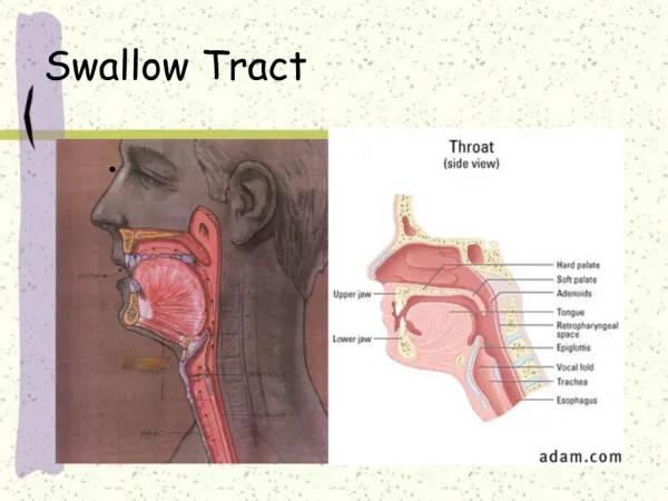
These problems can be detected with a barium swallow: • Narrowing of the esophagus (the muscular tube between the back of the throat and the stomach) • Disorders of swallowing • Abnormally enlarged veins in the esophagus that cause bleeding • Ulcers • Tumors • Polyps (growths that are usually not cancerous, but could be precancerous)
To Prepare: • Do not eat or drink anything, including water, after midnight before the test. • If you are pregnant, or think you might be, tell the staff before the x-ray is taken.
When the esophagus is empty , the mucosa coat is thrown into three or four straight longitudinal folds appeared as straight parallel lines throughout the esophagus
Normal indentations There are Two normal indentationsof the esophagus 1- At the aortic arch 2- At the left bronchus
Basic Positions • RAO (35° to 40°) • Lateral • AP • LAO
AP - LAT • AP
LAT - RAO • LAT
LAT - RAO • LAT
Lateral • Because the esophagus is thrown away from the spine, allowing better visualization
LAT - RAO • RAO
LAT - RAO • RAO
RAO • The esophagus is seen between the heart and the spine • The patient is rotate 35- 40 degrees with the RT side against the table
1.Oesophageal Varices • They appear inside the oesophagus and occasionally they occur in the stomach. Varices develop when most of the normal liver tissue has been replaced by scar tissue. Because the scar tissue pushes upon the veins in the liver, blood cannot flow normally through the veins.
What is the radiographic appearance ? • There are multiple submucosal filling defects in a barium filled esophagus in AP view.
2. Achalasia • Achalasia is a disease of esophagus . The term achalasia means "failure to relax" and refers to the inability of the lower esophageal sphincter (a ring of muscle situated between the lower esophagus and the stomach) to open and let food pass into the stomach. As a result, patients with achalasia have difficulty in swallowing food.
Lower Esophagus Proximal Dilatations Distal Narrowing
What is the radiographic appearance ? • Dilated smooth outlined barium filled esophagus with narrow tapering lower end of the esophagus with smooth outline and absence of fundal gas in stomach. . ( Rat Tail or Bird Beak Deformity)
3. Zenker'sdiverticulum (ZD) • Is a blind sac (pouch) that branches off the cervical esophagus. It is the most common type of esophageal diverticulum.
3. Zenker'sdiverticulum (ZD) • diverticula appeared as smoothly marginated round-to-ovoid sacs. The size of the openings of the diverticula depended on the location of the barium bolus .
4.Foreign body • Anteroposterior chest radiograph depicts a penny at the thoracic inlet of a 13-month-old infant who refused to eat.
4.Foreign body • Lateral chest radiograph depicts soft-tissue thickening at the tracheoesophageal interface. A penny was removed at esophagoscopy.